| المجلد 3 , العدد 9 , رمضان 1426 - تشرين الأول (أكتوبر) 2005 |
| |
| Tumor Necrosis Factor Alpha (TNFa?), Interleukin-1 Beta (IL-1?)
and Body Mass Index (BMI) in some of Hereditary Anaemia Diseases
|
| Epidemiologie de la Leishmaniose Cutanée en Syrie |
| د. علاء الشيخ حسن و د. فايزة القبيلي و د. غادة الأخرس |
| Al-Sheikh Hasan A., Al-Quobaili F. and Al-Akhras Gh. |
| كلية الصيدلة، جامعة دمشق - Faculty of pharmacy, Damascus University |
| Abstract |
هدفت هذه الدراسة إلى تحديد التبدلات في مستويات كل من عامل نخر الورم ألفا (TNFaa) والإنترلوكين -1 بيتا (IL-1b) عند مرضى بعض اضطرابات فقر الدم الوراثية والعلاقة المحتملة بين كل من الواصمين المشار إليهما أعلاه وبين منسب كتلة الجسم.
شملت الدراسة 113 مريضاً، 37 ثلاسيمية بيتا كبرى، 24 ثلاسيمية بيتا وسطى، 29 ثلاسيمية بيتا منجلية، 23 فقر الدم المنجلي و 24 شخصاً صحيحاً ظاهرياً كشواهد. تم قياس كل من TNFaa و IL-1b باستخدام المقايسة المناعية باللمعان الكيميائي.
كانت مستويات كل من TNFaa و IL-1b أعلى بشكل يُعتد به عند مجموعات المرضى أكثرمنها عند مجموعة الشاهد (P<0.05)، في حين كانت قيم منسب كتلة الجسم أخفض عند المرضى بالمقارنة مع الشواهد (P<0.05). كان ثمة ارتباط سالب يُعتد به بين قيم منسب كتلة الجسم وبين تراكيز كل من TNFaa (R=-0.31, P<0.05) و IL-1b (R=-0.25, P<0.05) عند جميع المرضى.
قد تساهم المستويات المرتفعة للـ TNFaa و IL-1b في مضاعفات اضطرابات فقر الدم الوراثية كمساهمة قيم منسب كتلة الجسم.
|
The aim of this study was to determine the changes in the level of Tumor Necrosis Factor Alpha (TNFaa) and Interleukin-1 Beta (IL-1b) in patients with hereditary anaemia disorders and the possible relationship between the above-mentioned markers and Body Mass Index (BMI).
The study included 113 patients of which, 37 have Beta Thalassaemia Major, 24 Beta Thalassaemia Intermedia, 29 Sickle-Beta+ Thalassaemia, 23 Sickle Cell Anaemia and 24 apparently healthy persons as controls. TNFaa and IL-1b were measured using chemiluminescence immunoassay.
Both TNFaa and IL-1b levels were significantly higher in patient groups than those of control group (P<0.05), while BMI values were lower in all patients in comparison to controls (P<0.05). There was a negative significant correlation between BMI values and both TNFaa (R=-0.31, P<0.05) and IL-1b (R=-0.25, P<0.05) concentrations in all patients.
Elevated levels of TNFab and IL-1b may contribute to complications of hereditary anaemia disorders such as BMI values.
الكلمات المفتاح Key words: عامل نخر الورم ألفاTumor Necrosis Factor Alpha، الإنترلوكين -1 بيتا Interleukin-1 Beta، منسب كتلة الجسمBody Mass Index، الثلاسيمية بيتا Beta thalassaemia، فقر الدم المنجليSickle Cell Anaemia.
|
| Introduction |
| Hereditary Anaemia Disorders are common in Syria. It is well known that Patients with hereditary anaemia disorders like Beta Thalassaemia and Sickle Cell Anaemia have increased morbidity and low perceived health status, similar to patients with chronic diseases. They suffer from many complications including wasting, Cachexia, and reduced body weight and body mass index (BMI), (1-3, 11).
Pro-inflammatory cytokines, such as Tumor Necrosis Factor alpha (TNFa) and interleukine-1 beta (IL-1b), may be involved in some of these complications due to various biological roles of the cytokines (4, 5).
It has been shown that elevated level of both cytokines play a major role in growth defect in patients with chronic diseases (1, 2, 6, 7).
|
| Materials and Methods |
Selection of Patients and Controls:
113 patients, aged 2-31 years, (11.5±5.7) were selected from patients referred to the National Center of Thalassaemia in Damascus. They were divided into four groups:
-Beta Thalassaemia Major: 37 Patients;
-Beta Thalassaemia Intermedia: 24 Patients;
-Sickle Beta+ Thalassaemia: 29 Patients;
-Sickle Cell Anaemia: 23 Patients.
Control group comprised 24 age-matched (2-28 years, 13.1±6.5) apparently healthy subjects. This group was randomly selected from a group of healthy population with no history of hereditary anaemia disorders and/or recent infection or inflammation.
Body Mass Index (BMI) values were calculated as follows:
BMI (Kg/m2) = Weight (Kg) / [height (m)]2.
A questionnaire about patients and controls age, sex and family history of hereditary disorders were collected.
Sampling: Blood samples were collected immediately before the date of periodic blood transfusion. Sera from patients and controls were separated and kept frozen at -20 C? for up to six months.
Measurement of TNFa and IL-1b levels: TNFa and IL-1b levels were measured using Chemi-luminescence Immunoassay using IMMULITE® analyzer (DPC, USA).
|
| Results |
Tables 1 and 2 & Figures 1 and 2 show the results of TNFa and IL-1b levels in patient and control groups.
BMI was calculated in all patients and controls, results are shown in table 3 and figure 3.
There was a negative significant correlation between BMI values and TNFa concentrations in all the patients (R = -0.31, P < 0.05) (Figure 4).
There was a negative significant correlation between BMI values and IL-1b concentrations in all patients (R = -0.25, P < 0.05) (Figure 5).
|
| Discussion and conclusion: |
The level of both TNFa and IL-1b levels were significantly higher in patient groups than those of control group. This elevation may be due to complications resulting from the accumulation of iron which damages most body organs. It is known that the defect in haemoglobin synthesis leads to increasing erythrocytes deterioration rate and iron overload with the resulting activation of mononuclear phagocytes by Interferon-Gamma (IFN?). However, there are other factors may be involved in this process such as platelets and placenta growth factor (PIGF) (2, 8). It has been noticed, in pervious studies of sickle cell anaemia, that there was an increase of monocytes percentage in peripheral blood, which express TNFa and IL-1b levels, from 0.3% to 0.4% in control and from 6.8% to 14.1% in patient’s groups. And it had showed that the peripheral blood monocytes of sickle cell anaemia patients had 139% and 34% more TNFa and IL-1b (respectively) per cell than normal monocytes (9,10).
Our results showed that the level of TNFa and IL-1b is higher in sickle cell anaemia and thalassaemia major patient groups than those of sickle-beta thalassaemia and thalassaemia intermedia patient groups. This may be attributed to the imbalance in the formation of haemoglobin chains is less in the later groups
than it is in the former ones.
BMI values were significantly lower in patient’s groups than those of control group (P<0.05).
|
Table 1: TNFa concentrations in patient and control groups.
Group |
N |
Range (pg/ml) |
Mean ± SD (pg/ml) |
Beta Thalassaemiaa Major |
24 |
0-21.5 |
8.3 ± 7.5 |
Beta Thalassaemiaa Intermedia |
13 |
0-18.4 |
7.8 ± 7.1 |
Sickle Beta+ Thalassaemiaa |
15 |
0-21.8 |
5.6 ± 6.1 |
Sickle Cell Anaemia |
13 |
0-25.1 |
8.6 ± 7.9 |
Control |
19 |
0-7.2 |
4.2 ± 1.9 |
N-number, SD- standard deviation |
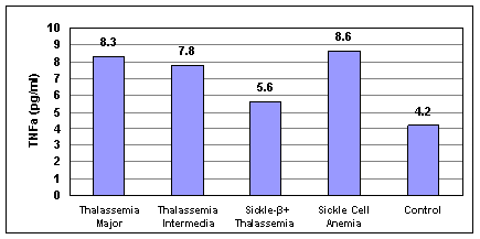
Figure 1: Mean concentrations of TNFa in patient and control groups.
Table 2: IL-1b concentrations in patient and control groups
Group |
N |
Range (pg/ml) |
Mean ± SD (pg/ml) |
Beta Thalassaemiaa Major |
37 |
0-32.7 |
6.6 ± 5.1 |
Beta Thalassaemiaa Intermedia |
11 |
0-33.9 |
5.3 ± 4.4 |
Sickle Beta+ Thalassaemiaa |
14 |
0-16.8 |
5.1 ± 4.7 |
Sickle Cell Anaemia |
10 |
0-18.6 |
7.5 ± 5.6 |
Control |
24 |
0-4.7 |
2.8 ± 1.6 |
N-number, SD- standard deviation |
|
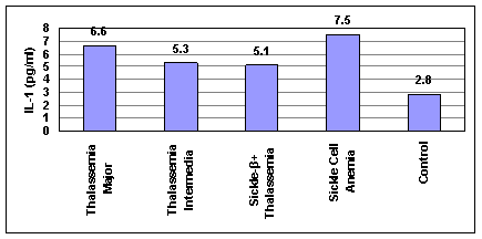
Figure 2: Mean concentrations of IL-1b in patient and control groups.
Table 3: BMI values in patient and control groups.
Group |
N |
Range (Kg/m2) |
Mean ± SD (Kg/m2) |
Beta Thalassaemiaa Major |
37 |
8.2-23 |
15.9 ± 2.4 |
Beta Thalassaemiaa Intermedia |
24 |
10.6-20.3 |
16.3 ± 2.8 |
Sickle Beta+ Thalassaemiaa |
29 |
11.5-24.2 |
16.5 ± 2.2 |
Sickle Cell Anaemia |
23 |
13.6-20.7 |
16.1 ± 1.9 |
Control |
24 |
17.4-25.7 |
20.7 ± 1.8 |
N-number, SD- standard deviation |
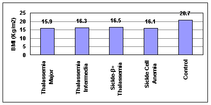
Figure 3: Mean values of BMI in patient and control groups.
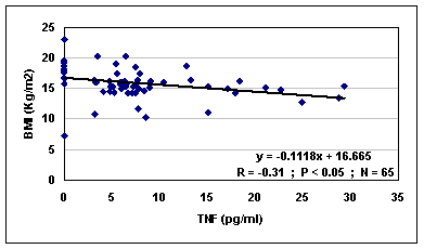
Figure 4: The correlation between TNFa and BMI in total patients group.
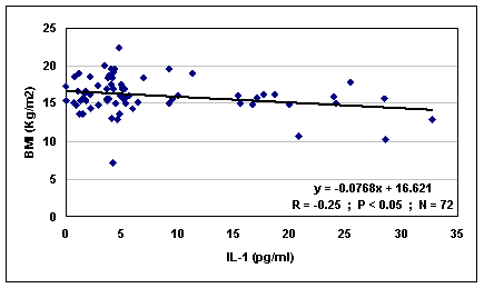
Figure 5: The correlation between IL-1b and BMI in total patients group. |
| There was a significant negative correlation between BMI values and the level of TNFa and IL-1b in all the patients.
It is known that TNFa induces wasting in human, which may explain why patients with high level of TNFa may suffer growth deficits (1). Furthermore, it is also known that IL-1b may affect the physical activity (6). Thus, high level of TNFa and IL-1b could contribute to complications of the disease such as cachexia (2).
TNFa and IL-1b measurements may be useful, in addition to other routine tests like WBC & Differential and Immunoglobulin Gamma (IgG) assay, in evaluating immunological state of patients of hereditary anaemia syndromes.
It should be stressed that there is a need for further study to explore possible relations between these markers and other markers such as IFN?, which may responsible for the complications of the immunological state in those patients, also the effects of these markers on the physical state of the patients.
|
| Acknowledgment: |
|
The authors would like to express their thanks and gratitude to Dr. Kusai AL Zir the manager of the National Center of Thalassaemia in Damascus and to all staff for their help and assistance, patients and persons who accepted to pose as control.
|
| References |
1-Kuvibidila S; Gardner R; Ode D; Yu L; Lane G. and Warrier RP.
Tumor Necrosis Factor alpha in children with sickle cell disease in stable condition.
J. Natl. Med. Assoc., 89(9): 609-615, 1997.
2-Wanachiwanawin W; Wiener E; Siripanyaphinyo U; Chinprasertsuk S; Mawas F. and Fucharoen S.
Serum levels of Tumor Necrosis Factor-alpha, interleukin-1, and interferon-gamma in beta (o)-thalassaemiaa/HbE and their clinical significance.
J. Interferon Cytokine Res. 19(2): 105-111, 1999.
3-Patey RA; Sylvester KP; Rafferty GF; Dick M. and Greenough A.
The importance of using ethnically appropriate reference ranges for growth assessment in sickle cell disease.
Arch Dis Child. 87(4): 352-353, 2002.
4-Francis RB Jr. and Haywood LJ.
Elevated immunoreactive Tumor Necrosis Factor and interleukin-1 in sickle cell disease.
J. Natl. Med. Assoc. 84(7): 611-615, 1992.
5-Santos-Rosa M; Bienvenu J. and Whicher J.
Cytokines. In: Tietz Textbook of Clinical Chemistry, 3rd ed. 21: 541-605, 1999.
6-Johansen KL; Kaysen GA; Young BS; Hung AM; da Silva M. and Chertow GM.
Longitudinal study of nutritional status, body composition, and physical function in hemodialysis patients.
Am. J. Clin. Nutr. 77(4): 842-846, 2003.
7-Cederholm T; Wretlind B; Hellstrom K; Andersson B; Engstrom L. and Brismar K.
Enhanced generation of interleukins 1 beta and 6 may contribute to the cachexia of chronic disease.
Am. J. Clin. Nutr. 65(3): 876-882, 1997.
8-Selvaraj SK; Giri RK; Perelman N; Johnson C; Malik P. and Karla VK.
Mechanism of monocyte activation and expression of proinflammatory cytochemokines by placenta growth factor.
Blood. 102(4): 1515-1524, 2003.
9-Wun T; Cordoba M; Rangaswami A; Cheung AW. And Paglieroni T.
Activated monocytes and platelet-monocyte aggregates in patients with sickle cell disease. Clin. Lab. Haematol., 24(2): 81-88, 2002.
10-Belcher JD; Marker PH; Weber JP; Hebbel RP. and Vercellotti GM.
Activated monocytes in sickle cell disease: potential role in the activation of vascular endothelium and vaso-occlusion. Blood, 96(7):2451-2459, 2000.
11-Srivatsa A. and Arivatsa A.
Assessment of adrenal endocrine function in Asian Thalassaemiacs.
Indian Pediatr. 7; 42(1): 31-35, 2005.
12-Rother RP; Bell L; Hillmen P. and Gladwin MT.
The clinical sequelae of intravascular hemolysis and extracellular plasma haemoglobin: a novel mechanism of human disease.
JAMA, 6; 293(13): 1653-1662, 2005.
|
| |
| المجلد 3 , العدد 9 , رمضان 1426 - تشرين الأول (أكتوبر) 2005 |
|
|
|
