| المجلد 4 ,
العدد 9
، ربيع الأول 1429 – نيسان (إبريل) 2008 |
| |
|
VIII Conference of the Syrian Clinical Laboratory Association |
| Four Seasons Hotel, Damascus |
| |
| Saturday 3 May 2008 |
H E Dr. Maher Housami,
Minister of Health |
Opening Ceremony
حفل الافتتاح |
09:30-10:15 |
Industrial Exhibition Opening
افتتاح المعرض العلمي |
10:15-10:45 |
Coffee Break 10:45 -11:15
Opening Lecture |
المحاضرة الافتتاحية |
Chair: Dr. Ghassan Shannan
Professor Eyad Chatty |
New Aspects in Medicine |
11:15-12:00 |
Plenary Lecture |
محاضرة رئيسية |
Chair: Dr. Emile Chahine
Dr. Dominique Kaiserlian |
CD8+ T cells as a novel therapeutic target in Crohn's disease |
12:00-12:30 |
|
| |
| CD8+ T cells as a novel therapeutic target in Crohn's disease |
Mucosal Immunology Group
INSERM-U851, IFR128, 21 Avenue Tony Garnier, 69365 Lyon CX 07, France |
| Crohn’s disease is a chronic T cell-mediated Inflammatory bowel diseases (IBD) whose ethiology and physiopathology is still unclear. Experimental evidence in both animal models and clinical studies in human have emphasized a unique role of Th1-type CD4+ T cells in active disease. Recently, we developed novel animal models, allowing to study the early phases of the mucosal immune response responsible for the acute phase of colitis as well as relapses of intestinal inflammation. We have shown that colitis results from a typical delayed-type hypersensitivity response responsible and that CD8+ T cells play a key role in the initiation of intestinal lesions by performing cytotoxicity against gut epithelial cells. Importantly, regulatory CD4+ T cells with suppressive function can inhibit in vivo priming of antigen-specific CD8 effector T cells and prevent susceptibility to colitis. Clinical studies in human Crohn’s disease carried out during the follow up of patients undergoing colon surgery allowed us to evaluate the prognosis value of increase in CD8 T cells expressing cytotoxic markers (granzyme B or perforine). Preliminary data revealed that an increased proportion of cytolytic CD8+ T cells in peripheral blood as well as in the mucosal lamina propria at the site of anastomosis, can be observed in patients before the relapse. These data illustrate that cytolytic CD8+ T cells are predictive marker of the relapse and represent a novel therapeutic target in Crohn’s disease .
|
| Saturday 3 May 2008 |
Symposium 1 Sequencing: Principles & Applications |
الجلسة العلمية الأولى |
Chairs: Dr. Sahar Fahoum, Dr. Wafeka Zaraour
Dr. Wafa Habal |
Principles of Sequencing: Overview |
12:30-13:00 |
|
DNA Sequencing: Principles & Applications
|
| Al-Assad Hospital, Damascus University, Damascus, Syria |
| DNA sequencing is the process of determining the nucleotide order of a given DNA fragment. In 1970s, two separate methods for sequencing DNA were developed: the chain termination method and the chemical degradation method. Both methods were equally popular to begin with, however, the former is more commonly used today. In this method, the DNA is used as a template to generate a set of labeled fragments that differ in length from each other by a single base. These fragments are then separated by size so that the bases and the original sequence are identified. The advent of DNA sequencing has significantly accelerated biological research and discovery. For the past decade, the output of sequence data from worldwide sequencing centers with constantly increasing sequencing capacities has been rising at an exponential rate. The rapid speed of sequencing attainable with modern DNA sequencing technology has been instrumental in the large-scale sequencing of the human genome as well as many animal, plant, and microbial genomes. The sequence of DNA encodes the necessary information for living organisms to survive and reproduce. Determining the DNA sequence, therefore, has been useful in basic research studying fundamental biological processes as well as in applied fields such as forensic and diagnostic research where it can be used to identify, diagnose and potentially develop treatments for genetic and infectious diseases. |
Dr. Wael El Garf |
DNA Re-sequencing |
13:00-13:30 |
|
| DNA Re-sequencing |
Wael Elgarf, M.D.
Biomedical Technology Department, National Research Center, Egypt
|
| Fluorescent DNA sequencing is rapidly becoming established as the technology of choice in basic research and clinical laboratories. The direct sequencing of an area of DNA using the product of a polymerase chain reaction (PCR) as template is a very powerful technique. It is straightforward and fast and therefore the method of choice in situations such as the analysis of mutations. The reliability of such analysis depends greatly on correct sequencing results. Automated DNA Sequencers generate a four-color chromatogram showing the results of the sequencing gel. They also generate the sample in the form of a text file of sequence data. The sequencer has no way of verifying the validity of the text data it generates. For the first few hundred nucleotides, the error rate may be low, but then errors occur more and more frequently as the resolution of the gel degrade. This is inherent in any sequencing gel, manual or automatic. Identifying the cause of a poor DNA sequencing result can often be very difficult as a particular sequencing problem may have many different underlying causes, or be the result of multiple interacting factors. Often the only way to work out the real cause of a particular problem is to perform a process of elimination. This process can be greatly simplified by visually examining both the raw and processed data chromatograms of the problematic sequencing traces. |
|
Discussion |
مناقشة |
13:30-13:45 |
Lunch Break 13:45 - 15:15
Workshop 1 |
جلسة العمل الأولى |
Chair: Dr. Tarek Dalati |
Distant Learning in Syria |
14:00-15:00 |
Symposium 2 Tuberculosis: Diagnosis & MDR-TB |
الجلسة العلمية الثانية |
Chairs: Dr. Mahmoud Zougheiby, Dr. El Taher Khaleel
Dr. Fadia Maamary |
Tuberculosis in Syria |
15:15-15:35 |
|
| ANTI-TUBERCULOSIS RESISTANCE IN SYRIA 2003 |
Syria: all Governorates
Study period febrile - October 2003
Dr Fadia Maamary
National Tuberculosis Control Programme, Ministry of Health, Damascus, Syria
|
This study Aimed at determine the first line anti TB drug resistance pattern among newly diagnosed sputum positive cases in Syria and Estimate the prevalence of the TB drug resistance among the previously treated sputum positive cases in Syria .
Design: The design chosen for implementing the survey was 100% sampling of diagnostic centers. The sample size was 304 new positive cases, and 72 previously treated the total period were 6 months.
Methods: Drug susceptibility testing to H, R, S, and E. was performed on Lowenstein-Jensen medium according to the proportion method.
Results: Of the (529) patient (120) sample were irregulars, 47 smears were negative, (362) smears were positive, (5) Cultures were contaminated, (5) cultures were negative (352) patients in analysis (295) with no history of previous anti-tuberculosis treatment and (57) with previous anti-tuberculosis treatment. The male female ratio was higher in "new" than in "previous treatment cases". Primary resistance to one or more drugs was found in 27.12% of new patients: Any resistance to H was (8.81%), any resistant to R was (5.76%), and any resistance to streptomycin was (23.39%). any resistance to Ethambutol was (5.42%). Multidrug resistance was (4.41%).
In previously treated patients were (57.89%) resistance to one or more drugs. Any resistance to H was (33.33 %) and any resistant to R was (29.82%) Any resistance to streptomycin was 31 (54.39%).any resistance to Ethambutol was (15.79%). Multidrug resistance was (24.56%).
Conclusions: Studies such as the should be conducted regularly to monitor drug resistance in Syria in order to effectively manage national tuberculosis control efforts.
|
Dr. D. Sarkis |
Lebanese Experience |
15:35-15:55 |
Dr. Faraj Barah |
Applications of Molecular Biology in Diagnosis and Management of Tuberculosis |
15:55-16:15 |
|
Applications of molecular biology in diagnosis and management of tuberculosis
تطبيقات البيولوجيا الجزيئية في تشخيص وتدبير داء السل
|
د. فرج بارة، كلية الصيدلة، جامعة القلمون الخاصة
Faraj Barah, College of Pharmacy, University of Kalamoon
|
Tuberculosis remains one of the biggest global health problems. One-third of the World’s population is latently infected with its causative agent; Mycobacterium tuberculosis. Since there is no treatment for some emerging multi-drug-resistant strains of Mycobacterium tuberculosis, there is a great concern that they may spread around the World. This emphasizes the need for additional control measures, such as new diagnostic tools, better drugs for treatment, and a more effective vaccine.
Laboratory diagnosis of tuberculosis currently relies on pulmonary symptoms, chest radiography, sputum smear microscopy and culture of the bacterial agent from clinical samples: all sharing various disadvantages. The tuberculin skin test is also commonly used to diagnose tuberculosis infection, but this test has poor specificity, poor sensitivity, cross-reactivity with many environmental Mycobacteria.
With the development of molecular biological techniques, methods of diagnosis of tuberculosis have been revolutionised, bringing an unprecedented opportunity for the rapid and specific detection of Mycobacterium tuberculosis, and overcoming the low sensitivity and specificity and long turnover time associated with traditional diagnostic tests. With automated sequencing analysis, species identification of Mycobacteria is now easier and more accurate than the conventional methods, and rapid detection of mutations, in the genes associated with the resistance of tuberculosis to drugs, provides early information on the potential drug resistance for each clinical isolate. However, the high cost of most of these new techniques, and their requirement for sophisticated equipment and skilled personnel have precluded their implementation on a routine basis, especially in low-income countries.
This presentation will focus on the most important current clinical applications of molecular biology in management of tuberculosis. Furthermore, the feasibility for their implementation in diagnostic laboratories will be discussed.
|
Dr. Asem Shehabi |
Recent Development of Antimicrobial Resistance in Mycobacteria Tuberculosis |
16:15-16:35 |
|
| Recent Development in Antimicrobial Resistance of Mycobacteria tuberculosis |
Asem A. Shehabi
Department of Pathlgy-Microbiology and Forensic Medicine, Faculty of Medicine, Unibversity of Jordan, Amman, Jordan.
|
Human Tuberculosis remains a major global health problem despite the presence of effective antituberculosis chemotherapy for over 50 years. Currently, there are more cases of TB in the world than ever recorded, particularly in developing countries. The massive reservoir of viable bacteria in the estimated 2 billion asymptomatically infected individuals worldwide causes serious problems for the control of TB. Multidrug-resistant tuberculosis (MDR-TB) has been recorded in 45 countries according to a new World Health Organization (WHO) report (2006). HIV infection contributes significantly to MDR-TB drugs. Selection for drug-resistant mutants in patients mainly occurs when patients are treated inappropriately or even are exposed to subtherapeutic drug levels. Over the past decade, much has been learned of the drug targets and mechanisms of resistance to first-line and several second-line antituberculosis agents. M.tuberculosis generally acquires drug resistance via de novon sSNP, a small deletions, or insertions in specific chromosomal loci, unlike most other pathogenic bacteria, which often acquire drug resistance via horizontal transfer. Molecular technique is can be easily used for detection and confirm of positive TB cases and to study the epidemiology of drug resistance, especially the prevalence and nature of genotype-specific mutations, and its association with phenotypic resistance . The threat created by TB drug resistance demands that a global and national plan should be implemented to reduce the incidence of TB in each country. |
|
Discussion |
مناقشة |
16:35-17:00 |
Exhibition Visit 17:00 - 18:00 |
| |
| Sunday 4 May 2008 |
Plenary Lecture |
محاضرة رئيسية |
Chair: Dr. Antoine Laham
Dr. Paul Wallace |
Flowcytometry: Overview |
09:00-09:45 |
Symposium 3
Flowcytometery: Principles & Applications |
الجلسة العلمية الثالثة |
Chairs: Dr. Fawza Monem, Dr. Hytham Kamal
Dr. Rabea Nahas |
The Diagnostic and Classification of Haematological Neoplasia by flow Cytometric Immunophenotyping |
09:45-10:15 |
|
تشخيص وتصنيف الخباثات الدموية بالتنميط المناعي بواسطة جهاز المقايسة الخلوية بالجريان
The diagnostic and classification of hematological neoplasias by flow cytometric immunophenotyping |
|
د. ربيعة النحاس اختصاصية في الطب المخبري – مشفى الأسد الجامعي بدمشق
|
• Flow cytometry is a rapid way to identify leukemias and lymphoma
• Abnormal antigen expression
• 98% correlation with Histopathology
• Can be used during treatment to monitor residual disease
The diagnostic and classification of hematological neoplasias by flow
cytometric immunophenotyping need a knowledge in:
– Clusters of differentiation (clusters designation) or CDs.
– Selection of reagents panels (monoclonal antibodies) and their fluorochromes.
– Staining.
– Data interpretation.
The final diagnostic classification of the disease is based on the immunological characterization of abnormal cells in conjunction with clinical findings, morphological and cytochemical and cytogenetic analysis and Bon Marrow study.
|
جهاز المقايسة الخلوية بالجريان مصطلح يطبق عملياً على الأجهزة التي تركز الضوء على الخلايا وتسجل تفلورها، والضوء المبعثر عنها. لنتمكن من قياس حجم الخلية ودراسة مواصفاتها
تعرف المقايسة الخلوية بالجريان أنه قياس الخلايا بنظام صمم ليوجه الخلايا في رتل واحد ضمن سائل حيث يتم تسليط أشعة الليزر عند نقطة القياس على الخلايا فيمر الضوء عبر هذه الخلايا ليتشتت 1- ما يسمى التشتت الأمامي FSC؛ 2- أو ينعكس بزاوية (تشتت جانبي SSC) ويقاس كلا التشتتين بواسطة كشافات ضوئية، كما يتم كشف تألق المواد الواسمة المتألقة والتي يتم ضبطها حسب طول موجة الليزر.
يرشح الضوء الصادر باستخدام مراشح ضوئية نوعية طول الموجة والتي تمرر أطوال موجة محددة وتعكس الأخرى.
يتم تحويل الضوء المنعكس إلى إشارة كهربائية بواسطة أنابيب مضخم ضوئي PMTS.
تفرز المعطيات ويتم تخطيطها بواسطة برنامج حاسوبي خاص وبذلك يمكن فرز أنماط مختلفة من الخلايا بدراسة ميزات المعطيات الناتجة وتكمن مقدرة
هذه التقنية في إمكانية قياس عدة معايير parameters في عشرات الآلاف من الخلايا المستقلة خلال عدة دقائق مما يسمح بتمييز الخلايا اعتمادا على صفاتها المستضدية والفيزيائية والكيميائية الحيوية.
تسمح هذه الطريقة بقياس عديد من المميزات لخلية مفردة.
1 - طريقة تسمح بقياس وكشف عدة صفات للخلية في آن واحد
Quantitative measure of multiple individual cellular properties:
2 - يتم هذا القياس أو الكشف أثناء مرور الخلايا (واحدة تلو الأخرى) في الجهاز الكاشف ضمن تيار من السائل، وبعد معالجة المستضدات الموجودة على سطح هذه الخلايا بأضداد وحيدة النسيلة موسومة بمادة مفلورة.
monoclonal antibodies labeled with fluorochromes dye and bound to WBC antigens
إذاً فإن جهاز الـ Flow Cytometer يستقصي كيفية تفاعل الخلية مع ضوء ليزري موجه عليها من خلال دراسة:
1- كيفية نثر الخلية للضوء الذي يعطي فكرة عن حجم الخلية وعن مدى تعقيدها الداخلي (التحبب).
- حجم الخلية: عن طريق قياس الضوء المنتثر المستقيم (forward scatter FSC).
- تحبب الخلية ومدى تعقيد محتواها الداخلي: عن طريق قياس الضوء المنتثر الجانبي (side scatter SSC).
2- كيفية ومدى تألق الخلية Fluorescence: بقياس كثافة تألقها بالفلورة FL1, FL2, FL3, FL4
وقد شملت تطبيقات القياس الخلوي بالتدفق، في العقدين الأخيرين، جميع فروع العلوم البيولوجية:
« إذ تمكننا هذه التقنية من:
- قياس خصائص عديدة للخلية في وقت واحد؛
- تحديد حجم الخلية؛
- تحديد درجة تحبب الخلية؛
- معرفة التعبير النوعي للمستضدات السطحية؛
- قياس التبدل في خصائص الخلايا من خلال قياس محتواها من الـDNA ؛
- كذلك قياس النشا ط الأنزيمي داخل الخلايا.
« تتحدد إمكانية هذه التقانة بالملونات المتألقة المستعملة، وخيال الباحث والذي غالباً ما يكون الأكثر أهمية.
The pathological cell population is identified by:
– Light scatter characteristics: uniform or abnormal.
– Abnormal antigen coexpression associated with different lineages.
– Asynchronous expression of antigens.
– Increased or decreased expression density of differentiation antigen.
– The relative expansion of cells of a single lineage.
– Selective expression of Kappa or lambda light chain (clonality).
– Abnormal maturation or reduction of mature elements of the same lineage.
Four patterns of antigen infidelity can be observed:
◊Aberrant expression of lineage-specific antigens.
◊ Asynchronous expression of lineage-associated antigens.
◊ Overexpression of cell surface antigens.
◊ Absence of cell surface antigen expression
-Immunophenotyping is useful in distinguishing acute myeloid leukemia (AML) from acute lymphoblastic leukemia (ALL) and further in classifying ALL into B-or T-cell lineage
◊ B-lymphoid markers: CD19, CD10 and cytoplasmic CD22 and CD79a
◊ T-lymphoid markers: CD2, CD7, (CD5) and cytoplasmic CD3.
◊ Myeloid markers: CD13, CD33, CD117 and cytoplasmic myeloperoxidase [anti-myeloperoxidase (MPO)]
◊ T – cell leukemias are relatively infrequent.
◊ Precursor – T ALL has a poorer prognosis than precursor – B ALL.
◊ Approximately 15% of childhood ALL are precursor – T ALL.
◊ Virtually all immature T-cell malignancies are TdT+ and CD7+. The lack of surface expression of CD3 is noticed in most cases of T-ALL.
◊ All subgroups of B-precursor ALL are
◊ TdT+ (except for mature B-All)
◊ Cyt CD79 a+
◊ Cyt CD22+
◊ CD19+
◊ Peripheral B-cell neoplasms:
1.B-CLL
2.Lymphoplasmocytoid lymphoma -immunocytoma
3.Mantle cell lymphoma
4.Hairy cell leukemia
5.Plasmacytoma/myeloma
6.Follicullar Center Cell (FCC)
grades:
I (small cell)
II (mixed small and large cell)
III (large cell)
7.Marginal zone B-cell
extranodal (MALT +/- monocytoid cells) nodal (+/- monocytoid cells)
splenic marginal zone (+/- villous lymphocytes)
8.Diffuse Large B-Cell include various subtypes , one defined: mediastinal (thymic) B-NHL
9.Burkitt´s lymphoma
10.High-grade Burkitt-like
Peripheral T cell and NK-cell neoplasms
1. T CLL
2. Large granular lymphocyte (LGL) leukemia T-cell type NK-cell type
3. Mycosis fungoides/Sezary syndrome
Peripheral T cell and NK-cell neoplasms
4. Peripheral T cell lymphoma cytologic categories: medium sized, mixed medium and large cell, large cell-lymphoepithelioid cell
5. Angioimmunoblastic T-cell lymphoma AILD
6. Angiocentric lymphoma
Peripheral T cell and NK-cell neoplasms
7. Intestinal T cell lymphoma (+/- enteropathy associated)
8. Adult T cell lymphoma/leukemia
9. Anaplastic large cell lymphoma CD30+, T-and null cell types
◊AML can be defined immunologically by the expression of two or more myeloid markers: CD13, CD33, CD117 and anti-MPO in the absence of lymphoid markers.
◊Combined use of CD13 + CD33 allows recognition of 99% of AML.
◊ The most specific marker for the myeloid lineage is anti-MPO followed by CD117, as a rule, both are negative in ALL |
Dr. Ibraheem Kebewar |
Application of Flowcytometry to sort myeloma cells for FISH analysis |
10:15-10:45 |
|
| Enrichment of plasma cells by sorting increases
the sensitivity of chromosomal abnormalities detecting in plasma cells myeloma in minimal residual diseases by FISH
|
Ibrahim Kebbewar1, AnneMarie W. Block1, Shelia N. Jani Sait1, George Deeb1, Petr Starostik1, Asher A. Chanan-Khan2, Paul K. Wallace1
Departments of 1Pathology and 2Medicine, Roswell Park Cancer Institute Buffalo, NY |
Plasma cell myeloma (PCM) is a monoclonal neoplasm of plasma cells that infiltrates the bone marrow with variable percentages. The detection of minimal residual disease (MRD) in PCM by fluorescence in-situ hybridization (FISH) has important diagnostic and prognostic implications. The cytogenetic studies indicated that 60-80% of patients with PCM have monoclonal IgH gene rearrangement detected in bone marrow samples. Based on our experience, bone marrow samples must contain at least 10% of plasma cells in order to detect chromosomal abnormalities by FISH. Plasma cells, however, are generally in very low frequencies in patient samples obtained after chemotherapy or bone marrow transplant. We found enriching the plasma cell pool using multi-parameter fluorescence activated cell sorting and subsequently performing FISH analysis on the sorted cells, increased the sensitivity of detecting the structural and numerical chromosomal abnormalities in plasma cells. Using this procedure, we could reliably detect chromosomal abnormalities in samples with as few as 0.5% abnormal cells. This methodology (Flow-FISH) is reliable, correlates flow cytometric phenotype with chromosomal genotype, and can be used to detect chromosomal abnormalities in minimal residual disease. |
Dr. Paul Wallace |
Clinical case studies and correlate these with histology, cytogenetic and molecular |
10:45-11:15 |
|
Discussion |
مناقشة |
11:15-11:30 |
Coffee Break 11:30 -12:00
Symposium 4 Homocystein |
الجلسة العلمية الرابعة |
Chairs: Dr. Wolfgang Herrmann, Dr. Muhelddin Jumaa
Dr. Wolfgang Herrmann |
Homocysteine a risk factor with higher age – vessel disease and osteoporosis |
12:00-12:30 |
|
| Homocysteine a risk factor with higher age-vessel disease and osteoporosis |
Wolfgang Herrmann
Department for Clinical Chemistry and Laboratory Medicine / Central Laboratory,
Medical School, Saarland University, D-66421 Homburg, |
The role of hyperhomocysteinemia (HHcy) as risk factor of cardiovascular diseases (CVD) is presently under debate. Despite the strong evidence for a causal relationship between HHcy and CVD, an open question in the causality issue remains; can HCY-lowering treatment reduce the incidence of CVD? Worldwide about 52,000 people are currently included in various prevention trials for addressing this issue (1). Few secondary intervention trials are meanwhile completed, VISP (3860 stroke patients were supplemented B-vitamins for 2 years) (2), NORVIT (3749 patients with MI received B-vitamins for 3 years) (3), and HOPE-2 (5522 patients with CVD or diabetes were treated with B-vitamins for 5 years) (4). These trials failed, however, to show an improvement in cardiovascular end points. Secondary prevention trials, however, have many limitations. Patients with CVD receive various medications which makes it difficult to find an additional or an independent effect for the vitamins. A very large number of CVD patients have to be included in randomised trials in order to demonstrate vitamin effects. For a risk reduction of 10% resp. 20% the number of study participants must be at least 60,000 resp. 12,000 (5). Another serious limitation is the insufficient duration of these trials to assure statistically valid results. A prospective meta-analysis of the ongoing HCY-lowering trials which include about 50,000 patients will be available in some years from now.
However, a very recent meta-analysis by Wang et al. (6) has contributed a lot in clarifying this inconclusive situation. This meta-analysis included eight randomised trials with about 16,841 patients provided convincing evidence that lowering of plasma HCY by folic acid supplementation reduces the stroke risk significantly by approximately 18%. The reduction of stroke risk was even higher when the treatment exceeded 36 months (-29%), the HCY lowering was more than 20% (-23%), the patients had no stroke history (-25%) or the patients consumed no folic acid enriched grain products (-25%). The authors concluded that folic acid supplementation can effectively reduce the risk of stroke in primary and secondary prevention. This is in line with results from the USA and Canada where food fortification with folic acid since 1998 has contributed to a decreased stroke risk (7). Since 1998, annually 13,000 less stroke death have been registered in the US. The meta-analysis by Wang et al. (6) also underlines the role of the duration of treatment for a successful risk reduction. Only intervention studies lasting longer than three years have responded with a significant lowering of stroke risk. Furthermore, the risk reduction in stroke by B-vitamins seems to be greater than in CVD.
Another important current issue in HCY research deals with chronic heart failure (CHF). CHF is a major public health problem causing considerable morbidity and mortality. In the elderly population >75 years the prevalence of CHF exceeds 10%. Recently, plasma HCY has been suggested to be increased in CHF patients potentially representing an additional important risk factor of CHF (8). Experimental HHcy in rats caused increased BNP concentration in myocardial tissue but also higher incidence of elevated plasma BNP level (9). In superfusion experiments utilising myocardial tissue rising HCY concentrations in superfusion medium caused increasing BNP excretion (9). The available data support a causal relationship between HHcy and CHF and suggest that HHcy contributes to adverse cardiac remodelling characterized by interstitial and perivascular fibrosis resulting in increased myocardial stiffness. In addition, HHcy seems to affect the pump function of the myocardium. Therefore, HHcy might be an important aetiological factor in CHF.
A further important aspect of HCY research is the relationship between HHcy and osteoporosis (10). Elderly people are the most important risk group and identifying this risk factor might have marked clinical implications. Prospective clinical trials have linked high HCY plasma levels and low B-vitamin concentrations in adults with an increased risk of fragility fractures and osteoporosis (11;12). The special interest in HHcy as risk factor for osteoporosis is on the one hand, due to its high prevalence in elderly people and on the other hand, HCY can easily be modified by B-vitamin supplementation. Experimental HHcy in rats has confirmed a stimulated bone resorption and accumulation of HCY in bone tissue by collagen binding (13). In vitro experiments showed that HCY and low B-vitamins levels stimulated osteoclast activity and caused a shift towards bone resorption. HHcy (and possibly B-vitamin deficiencies) adversely affects bone quality by a stimulation of bone resorption and disturbance of collagen cross linking. Thus, a causal involvement of HHcy in osteoporosis is suggested.
Because HHcy is very common in elderly people and its prevalence is growing due to our rapidly aging population, it seems important to identify subjects with vitamin deficiency and to ensure sufficient vitamin intake for primary and secondary prevention of degenerative age related diseases.
Reference List
1. B-Vitamin Treatment Trialists' Collaboration. Homocysteine-lowering trials for prevention of vascular disease: protocol for a collaborative metaanalysis. Clin Chem Lab Med 2007; 45:1575-81.
2. Toole JF, Malinow MR, Chambless LE, Spence JD, Pettigrew LC, Howard VJ, et al. Lowering homocysteine in patients with ischemic stroke to prevent recurrent stroke, myocardial infarction, and death: the Vitamin Intervention for Stroke Prevention (VISP) randomized controlled trial. JAMA 2004; 291:565-75.
3. Bonaa KH, Njolstad I, Ueland PM, Schirmer H, Tverdal A, Steigen T, et al. Homocysteine lowering and cardiovascular events after acute myocardial infarction. N Engl J Med 2006; 354:1578-88.
4. Lonn E, Yusuf S, Arnold MJ, Sheridan P, Pogue J, Micks M, et al. Homocysteine lowering with folic acid and B vitamins in vascular disease. N Engl J Med 2006; 354:1567-77.
5. Wald DS. Folic acid, homocysteine and cardiovascular disease - judging causality in the face of inconclusive trial evidence. World Congress on Hyperhomocysteinemia. Clin Chem Lab Med 2007; 45(5):A17.
6. Wang X, Qin X, Demirtas H, Li J, Mao G, Huo Y, et al. Efficacy of folic acid supplementation in stroke prevention: a meta-analysis. Lancet 2007; 369:1876-82.
7. Yang Q, Botto LD, Erickson JD, Berry RJ, Sambell C, Johansen H, et al. Improvement in stroke mortality in Canada and the United States, 1990 to 2002. Circulation 2006; 113:1335-43.
8. Vasan RS, Beiser A, D'Agostino RB, Levy D, Selhub J, Jacques PF, et al. Plasma homocysteine and risk for congestive heart failure in adults without prior myocardial infarction. JAMA 2003; 289:1251-7.
9. Herrmann M, Taban-Shoma O, Hubner U, Pexa A, Kilter H, Umanskaya N, et al. Hyperhomocysteinemia and myocardial expression of brain natriuretic peptide in rats. Clin Chem 2007; 53:773-80.
10. Herrmann M, Schmidt JP, Umanskaya N, Wagner A, Taban-Shomal O, Herrmann W. The role of hyperhomocysteinemia and B-vitamin deficiencies in osteoporosis - a systematic review. Clin Chem Lab Med 2007; 45:in press.
11. van Meurs JB, Dhonukshe-Rutten RA, Pluijm SM, van der Klift M, de Jonge R, Lindemans J, et al. Homocysteine levels and the risk of osteoporotic fracture. N Engl J Med 2004; 350:2033-41.
12. McLean RR, Jacques PF, Selhub J, Tucker KL, Samelson EJ, Broe KE, et al. Homocysteine as a predictive factor for hip fracture in older persons. N Engl J Med 2004; 350:2042-9.
13. Herrmann M, Wildemann B, Claes L, Klohs S, Ohnmacht M, Taban-Shomal O, et al. Experimental Hyperhomocysteinemia Reduces Bone Quality in Rats. Clin Chem 2007; 53:1455-61.
|
Dr. Rima Obaid |
Methylation and neurodegeneration |
12:30-13:00 |
|
| Methylation and neurodegeneration |
|
Rima Obeid1, Mariz Kasoha1, Jean-Pierre Knapp1, Panagiotis Kostopoulos 2, Klaus Fassbender 2, Wolfgang Herrmann1*
1Department of Clinical Chemistry and Laboratory Medicine, 2Department of Neurology, Faculty of Medicine, University Hospital of Saarland, Homburg/Saar |
| Background: B-vitamins (folate, B12) are important micronutrients for brain function and for homocysteine (Hcy) metabolism. Increased plasma concentration of total Hcy (tHcy) has been related to dementia, but the mechanisms have not been well investigated. We studied the role of the B-vitamins in tHcy metabolism in the brain.
Methods; We studied blood and cerebrospinal fluid (CSF) samples from 182 patients. We measured tHcy, cystathionine, S-adenosyl methionine (SAM), S-adenosyl homocysteine (SAH), and the B-vitamins. In addition, we measured concentrations of phosphorylated tau protein (181P) (P-tau) and b-amyloid(1-42) in the CSF.
Results: Concentrations were lower in CSF than serum or plasma for tHcy (0.09 vs. 9.4 umol/l), SAH (13.2 vs. 16.8 nmol/l), cystathionine (54 vs. 329 nmol/l), and holotranscobalamin (16 vs. 63 pmol/l), whereas concentrations in CSF were higher for SAM (270 vs. 113 nmol/l) (all p <0.05). CSF tHcy was predicted by concentrations of CSF cystathionine (b=0.478), folate (b=-0.403), albumin (b=0.349), and age (b=0.298). Additionally, aging was associated with higher concentrations of SAH in the CSF, lower concentrations of CSF folate and lower SAM/SAH ratio. We found at lower serum folate a higher CSF-folate compared to serum folate. This relationship was in contrast to that found at high serum folate. Concentrations of CSF SAH and CSF folate correlated significantly with those of P-tau (r=0.46 and r=-0.28, respectively). Moreover, P-tau correlated negatively with SAM/SAH ratio (r=-pendent on age.
Conclusions: Age is associated with higher CSF-tHcy and CSF-SAH and lower CSF-folate. Elevation of CSF-SAH was related to a higher CSF-P-tau that might be related to a decreased degradation of P-tau. Disturbed methyl group metabolism may be the link between hyperhomocysteinemia and neurodegeneration. We anticipate that lowering tHcy and SAH might protect the brain by preventing P-tau accumulation.
|
Dr. Abdul-Rahman Al-Bazzaz |
Is there any advantage to the measurement of S-adenosylhomocysteine rather than homocysteine as a cause of the vascular damage and its mechanism of action? |
13:00-13:30 |
|
| Is there any advantage to the measurement of S-adenosylhomocysteine rather than homocysteine as a cause of the vascular damage and its mechanism of action? (Review) |
Abdul-Rahman A. Al-Bazzaz
Al-Nahrain University, College of Medicine, Baghdad, Iraq.
Now : Al-Ahliyya Amman University, Faculty of Pharmacy and Medical Sciences. Amman- Jordan
|
Abstract: Chronic heart failure (CHF) is a major public health problem causing considerable morbidity and mortality. Prevention of CHF by identifying risk factors is therefore a major issue. Previous studies found that hypertension, smoking, diabetes mellitus, obesity, and advancing age are the most important risk factors for CHF. Recently, plasma homocysteine (Hcy) has been suggested as a newly recognized risk factor. However, there are no data regarding the association between Hcy and various objective as well as subjective measures of CHF.
It is widely accepted that elevated plasma total homocysteine is an independent risk factor for vascular disease. The relation is believed to be causal, but there is no generally accepted mechanism for the pathophysiology involved. The metabolic precursor of homocysteine in all tissues is S-adenosylhomocysteine (AdoHcy). AdoHcy is present in normal human plasma at concentrations approximately 1 - 500th of those of homocysteine, a fact that presents difficulties in measurement. The requirement for specialized equipment, complicated time-consuming methodology, or both is a reason that measurement of plasma AdoHcy has not generally been carried out in large studies. A recently published rapid immunoassay for AdoHcy in human plasma should make measurement of this important metabolite available for general use. Advantages of the measurement of plasma AdoHcy include 1) a smaller overlap of values between control subjects and patients, and thus the possibility of observing significant differences in fewer samples, 2) an accepted mechanism of metabolic activity as an inhibitor of all S-adenosylmethionine–mediated methyltransferases, and 3) evidence (from recent studies) that a higher plasma concentration of AdoHcy is a more sensitive indicator of vascular disease than is a higher plasma concentration of homocysteine.
Several small studies have shown that measurement of plasma AdoHcy is a better indicator of the risk of vascular disease than is measurement of plasma tHcy. Studies firstly showed that, when compared with control subjects, patients with end-stage renal disease had 44-fold greater plasma AdoHcy but only 5-fold greater plasma homocysteine concentrations. Both measurements were significantly (P < 0.001) different, but the authors drew no conclusions about which measurement was more sensitive. In a study published in 2001 comparing patients with proven CVD and matched controls, there was a significant difference in the plasma AdoHcy concentrations between the patients and controls but no significant difference in the homocysteine concentrations.
There is no generally accepted mechanism for the pathophysiology of elevated plasma homocysteine as a cause of vascular disease. Various mechanisms for the toxic action of homocysteine include a change in the redox status of the tissues with production of reactive oxygen species; an inhibition of anticoagulation mechanisms mediated by the vascular endothelium; antiplatelet effects related to the reaction of elevated homocysteine with nitric oxide to form S-nitrosohomocysteine; a direct effect of homocysteine on vascular endothelial or smooth muscle cells; the formation of homocysteine thiolactone that modifies endothelial proteins; and the induction of programmed death of endothelial cells. In most cases, these actions have been indicated by effects caused by the addition of homocysteine to cells in culture. The principal problem with most of these studies has been the use of concentrations of homocysteine far higher (50–1000 µmol/L) than those present in plasma to show these effects. Rarely have any effects been shown with concentrations of homocysteine as low as 10 µmol/L. Homocysteine has a free sulfhydryl group and is oxidized with a second homocysteine molecule to form the disulfide, homocystine, and also with cysteine to form a mixed disulfide. In human plasma, most homocysteine exists in disulfide linkage to cysteine in albumin. For this reason, it has been the standard practice to measure total homocysteine (tHcy) that is produced after the reduction of the bound homocysteine. The normal concentration of tHcy in human males is 10µmol/L. However, the amount of free homocysteine in human plasma is <1% (< 0.1 µmol/L). Therefore, although high plasma concentrations of homocysteine are associated with vascular disease, it has been difficult to show
that they are the proximal cause of the damage.
An alternative possible cause of the pathophysiology associated with hyperhomocysteinemia is AdoHcy. This compound is the precursor of all of the homocysteine in tissues. Except for methyl transfer from betaine and from methylcobalamin in the methionine synthase reaction, AdoHcy is the product of all methylation reactions that involve S-adenosylmethionine (AdoMet) as the methyl donor. There are 50 reactions that carry out methyl transfer in cells. AdoHcy is well known as a potent inhibitor of most, if not all, methyltransferases. Increased concentrations of AdoHcy in tissues are usually accompanied by decreased concentrations of AdoMet. The use of the ratio of AdoMet to AdoHcy as an indicator of the methylating capacity of the cell was first suggested by Cantoni et al, and this ratio has been referred to as the "methylation index". However, in certain situations, the elevation of AdoHcy appears to be a better indication of the inhibition of methylation than does the ratio of AdoMet to AdoHcy. Methylation is significant in epigenetic regulation of protein expression via DNA and histone methylation. The inhibition of these AdoMet-mediated processes by AdoHcy is a proven mechanism for metabolic alteration. Because the conversion of AdoHcy to homocysteine is reversible, with the equilibrium favoring the formation of AdoHcy, increases in plasma homocysteine are accompanied by an elevation of AdoHcy in most cases.
|
|
Discussion |
مناقشة |
13:30-13:45 |
Lunch Break 13:45 - 15:15
Sunday 4 May 2008
Workshop 2 |
جلسة العمل الثانية |
Chair: Dr. Eyad Tenbakji |
Quality Management in Syria |
14:00-15:00 |
Symposium 5 Accreditation |
الجلسة العلمية الخامسة |
Chairs: Dr. Adel Noufal, Dr. Naheed Bashour
Dr. Ghassan Shannan |
The Time is near for Laboratory Accreditation in Syria |
15:15-15:30 |
Dr. Youssef Bilto |
Laboratory accreditation initiatives in neighbouring countries |
15:30-15:45 |
Dr. Ken Sikaris |
The importance of professional organisations in laboratory accreditation |
15:45-16:05 |
Dr. Elizabeth Frank |
How to manage an accredited laboratory |
16:05-16:30 |
Dr. Ken Sikaris |
Does accreditation improve analytical quality |
16:30-16:45 |
|
Discussion |
مناقشة |
16:45-17:00 |
Exhibition Visit 17:00 - 18:00
Monday 5 May 2008
Plenary Lecture |
محاضرة رئيسية |
Chair: Dr. Abdulaghani Maa El Bared
Dr. Mathias Montenarh |
Cancer Treatment: cell cycle interferences and apoptosis induction |
09:00-09:45 |
Symposium 6 Molecular Biology |
الجلسة العلمية السادسة |
Chairs: Dr. Mouhamed Tinawi, Dr. Ghada Akhrass
Dr. Maurizio Ferrari |
New trends in molecular diagnostics |
09:45-10:15 |
Dr. Mariam Klouche |
Molecular diagnosis of infectious diseases |
10:15-10:45 |
|
| Molecular diagnosis of infectious diseases |
|
Mariam Klouche
|
| Molecular diagnosis of infections has considerably improved sensitivity, specificity and speed of detection of many pathogens. Besides identification of acute or latent infection, quantitative molecular testing has a central role in monitoring of treatment. For several viral infections, quantification of the viral load forms part of prognostic algorithms, allowing individually adapted treatment with improvement of patient outcome. In fact, the introduction of systematic PCR-testing for HIV, HCV and HBV in blood donors in Europe was the single most important measure to reduce transfusion-associated transmission of infections. Since not even 10 years ago, initial viral genotypic resistance tests became available, allowing the prediction of susceptibility prior to antiviral or antiretroviral treatment. Even more recently, direct identification technologies for pathogenic microorganisms are emerging to be applied in diagnosis of serious bloodstream infections and infections at other sterile body sites. Compared to the rather slow traditional microbiologic tests, rapid bacterial or fungal nucleic acid-based technologies aim at identification of pathogenic micoorganisms and conceivably antimicrobial resistance within minutes to hours. Interpretation of direct detection of panbacterial or panfungal nucleic acids instead of living micoorganisms in blood is complex, given the risk of contamination, the ubiquitous presence of bacterial and fungal DNA, and the lack of a gold standard. Standardisation remains an important issue for several molecular diagnostic applications in infectious diseases. This presentation summarises the current applications of molecular diagnosis of pathogenic microorganisms and highlights the future perspectives with a particular focus on standardisation and the potential clinical usefulness in infectious disease diagnosis. |
Dr. Batool Shannan |
Molecular approaches to study predisposition to diseases |
10:45-11:15 |
|
Discussion |
مناقشة |
11:15-11:30 |
Coffee Break 11:30 -12:00 |
| |
| Monday 5 May 2008 |
| |
Symposium 7 FISH |
الجلسة العلمية السابعة |
Chairs: Dr. Halema El Alami, Dr. Mahjoub Jairoudi
Dr. Mazen Kanj |
Leukemia: Cryptogenic Approach |
12:00-12:30 |
Dr. Said Hammad Abdou |
Basics and Clinical Applications of Fluorescence in Situ Hybridization, FISH |
12:30-12:55 |
|
| 32- Basics and Clinical Applications of Fluorescence in Situ Hybridization (Fish) |
|
Said Hammad Abdou
Molecular Biology Unit, Tanta Universityِِ, Egypt.
|
During the last 15 years, molecular biology methods expanded into human cytogenetics. This is in close connection with advanced technologies of DNA probes preparation and possibilities of their non-radioactive labeling by means of enzymatic incorporation of modified nucleotides and their hybridization with complementary DNA of chromosomes and interphase nuclei.
FISH based on the unique ability of a portion of single stranded DNA, i.e., a probe, to anneal with its complementary target sequence wherever it is located on a metaphase spread. Thus, a DNA probe for a specific chromosomal region will recognize and hybridize to its complementary sequence on a metaphase chromosome or within an interphase nucleus. Both have to be in single-strand conformation, therefore, the DNA probe and the target DNA must be denatured, usually by heating them in Formamide-containing solution.
The probe is hybridized to the target DNA under conditions that allow the DNA to reanneal in double-stranded form. The probe DNA can be observed on its target by using a fluorescent microscope with filters specific for the fluorochrome label and the counter stain. Special dual and triple-pass filters have been developed to allow simultaneous visualization of several fluorohromes. Digital cameras designed to detect low light level emissions and computer imaging are used to increase the sensitivity of probe detection. Because fluorescent dyes are subjected to photo-bleaching (fading), the preparations are not permanent and must be stored away from light. Use of an anti-fade solution (phenylene-dianine) has improved the capacity to observe and document fluorescently labeled samples. Unlike most other methods used for chromosome analysis, FISH can be used to study chromosomes, which are resting between cell division in the interval known as interphase. Hence, this study is sometimes referred to as interphase cytogenetics. In this lecture, we will highlight on basics of FISH technique, Clinical applications e.g. Diagnosis of hematological malignancies, follow-up & detection of minimal residual disease, microdeletion syndromes and prenatal diagnosis.
|
Dr. Kamel Kebbewar |
Karyotyping and its Application in the Diagnosis of Trisomiers and other Chromosomal Abnormalities |
12:55-13:15 |
Dr. Walid Daass |
Finger Printing |
|
13:15-13:35 |
|
Discussion |
مناقشة |
13:35-14:00 |
|
Closing Remarks |
|
14:00-14:15 |
Lunch Break 14:15 - 16:00
Exhibition Visit 16:00 - 18:00 |
| 1- تجربة رائدة لهيئة المخابر في سورية - تدريب/ تعليم عن بعد |
|
د. حسن السيد و د. غسان شنان
|
أصبح التعليم عن بعد (Distance Learning) نهجاً حضارياً مرموقاً ومنتشراً في عدد كبير من المؤسسات التعليمية في العالم، بعد التقدم الباهر الذي شهدته الانترنيت (الشابكة) وما يتعلق بها من علوم المعلوماتية والمعدات المخدِّمة لها بما في ذلك شبكة الاتصالات الخاصة (Public Data Network-PDN) والشبكة اللاسلكية (Wi-Fi) وأخيراً الشبكة الخليوية (cellular networks).
تقوم الآن هيئة المخابر الطبية في سورية (بالتعاون مع الاتحاد الدولي للكيمياء السريرية IFCC
ومؤسسة تعليمية محلية)، بتجربة أولية في التعليم عن بعد على شكل دورة تدريبية لمجموعة مختارة من اختصاصيي المخابر الطبية في محافظات سورية.
تم اختيار "أساسيات الجودة الداخلية في المخبر الطبي" كموضوع للدورة التدريبية نظراً لأهمية الموضوع وسهولة تناوله من قبل المدربين والمتدربين. كما تم اختيار المتدربين على أساس مقدار معرفتهم باستعمال الحاسوب والانترنيت واللغة الانكليزية.
اختير استعمال "نهج التعليم عن بعد" في الدورة في مرحلة التحضير النظري للمتدربين من حيث توضيح الأسس النظرية للجودة الداخلية، وطُعِّم ذلك بنهج التدريب التقليدي في مرحلة التطبيق العملي لمفاهيم الجودة الداخلية في المخبر.
تأمل الهيئة في تحسين هذه التجربة وتطويرها كي تكون نموذجاً يعتمده ويدعمه الاتحاد الدولي، ويتم تعميمه على عدد من الدول المجاورة في إقليم شرق البحر المتوسط وفي أقاليم أخرى من العالم.
|
2- إطالة العمر بين الخيال العلمي والواقع
Theories about Aging and Mechanism of Anti – Aging |
|
د. رحاب الصّواف
|
لن يرضى الباحثون في مجال طب الألفية الثالثة بمعالجة العلل والأمراض فحسب، بل سيهدفون إلى إيجاد طريقة لخداع الموت ذاته، خاصة بعد أن أصبحوا الآن أقرب إلى فهم الآليات الإمراضية المتعلقة بالشيخوخة والمؤدية إلى الموت.
دارت الأيام وازداد الخيال العلمي توسعاً وتحليقاً ضمن مجالات عدة، منها الأبحاث الوراثية (الجينية)
التي طالت العديد من المواضيع المباحة وغير المباحة وعلى رأسها الشيخوخة وطرق الهروب منها أو تأخيرها سواءً بأساليب علمية أو خيالية نحن بانتظار نتائجها وتداعياتها.
وقد حاول البعض تحليل أسباب الشيخوخة والعوامل المؤثرة فيها من أمراض أو أسباب بيئية يمكن التحكم فيها وخلق توازن جديد في العضوية يعيدها إلى نشأتها.
يستهتر الإنسان اليوم بقدرته على المحافظة على شبابه، فهو يأكل بنهم، ويتناول الأدوية بشكلٍ فوضوي ويدخن السجائر ويرمي الأكاسيد والفضلات في الهواء وفي الماء وفي التراب حتى أصبح العالم بؤرة للملوثات البيئية والجرثومية والتي بلا شك تعتبر من أهم أسباب الشيخوخة التي كان باستطاعته ضبطها والحد منها بأساليب عدة.
قال الله تعالى: (لقد خلقناكم في أحسن تقويم) صدق الله العظيم.
وعلى هذا ظهرت نظريات عدة تناولت طرق المحافظة على الشباب وإطالة العمر نذكر منها:
النظريات الوراثية:
تعتمد على عدة طرق منها:
1. التداخل على المادة الوراثية لدى الإنسان، وقد بدأت تطبق فعلياً على الحيوانات ( فئران - ديدان مستديرة – ذبابة الفواكه) حيث استطاعوا إطالة عمر الفئران بنسبة 30% زيادة عن العمر الطبيعي.
2. المعالجة الوراثية التي تعتمد على تعديل مورثة الشيخوخة (المكتشفة على الصبغي الرابع).
3. استعادة قدرة الإنسان على المحافظة على شبابه عن طريق استعادة الهرمون المفقود لديه ألا وهو الفيتامين C الذي يعتقد أن أجدادنا كانوا يملكونه وقد فقد مع الزمن بسبب حدوث طفرات على المادة الوراثية للإنسان، فجميع الثديات تمتلك مورثات طبيعية مسؤولة عن تصنيع الفيتامين C اعتباراً من الغلوكوز باستثناء الثديات العليا (الإنسان والقرود(.
4. تثبيت المُتَقَدِّرات (Mitochondria the Fix) والحفاظ على سوية عملها بشكل جيد حيث يعتقد العلماء أن أي خلل في وظيفتها يؤدي إلى خلل مرضي في العضوية مثل السكري وإلزهايمر وآلام المفاصل وغيرها. فبتثبيت المُتَقَدِّرات نستطيع تثبيت العمر الخلوي ومنه إطالة العمر.
5. التداخل على بروتين التليمير telemer الموجود في الخلية الحية وهو جزء من الـ DNA المسؤول عن الانقسام الخلوي والذي يمكن أن يمنح الخلود للخلايا في حال تطبيق تقنيات الهندسة الوراثية عليه شرط أن لا يؤدي ذلك إلى حدوث أورام نتيجة الإنقسام اللانهائي.
كما ظهرت نظريات عديدة أخرى طالت نفس الهدف لكنها مازالت ضمن الخيال العلمي والذي
سيبقى ضمن هذا الإطار إذا نظرنا على مستوى الغد القريب.
بانتظار نتائج هذه الأبحاث هل سنبقى مكتوفي الأيدي لنعرف مدى نجاحها أو فشلها أم علينا التحرك سريعاً بهدف تأخير أعراض الشيخوخة والمحافظة على الشباب قدر الإمكان فنحن لا نريد زيادة العمر على حساب الصحة.
يجب على طب المستقبل أن يركز على النقاط التالية الممكن تحقيقها فعلياً على المستوى القريب أكثر من الحلم بها مثال ذلك:
1) مراقبة تناول الأدوية الذي يتم بشكل عشوائي، حيث يعتبر تناول الدواء بشكل خاطئ هو السبب الرابع للوفيات في الولايات المتحدة الأمريكية .
وعلى هذا فإن زَبْوَنِة الدواء Customized medicine أي إعطاء الدواء بحسب الزبون هو خيال علمي أصبح علماً فعلى المرء تصوير مورثاته قبل تناول أي دواء وذلك للتأكد من ملائمته له.
وهناك اختبارات وراثية بُدء بتطبيقها ضمن مجال الخريطة الوراثية Genes mapping كاختبارات الـ:
1. Liver Genomics
2. Neuro Genomics
3. Cardiac Genomics
4. Cancer Genomics
2) إعادة توازن الهرمونات التي تختل مع التقدم بالعمر (هرمون النمو- بروجسترون - تستسترون ... )
3) ضبط الداء السكري والعوامل المؤهبة له والذي ازداد تواتره في العالم مع زيادة البدانة والعادات الغذائية الخاطئة حيث قاربت نسبته الـ 20% خاصة في دول الخليج العربي.
4) خفض نسبة المدخنين بإتباع الوسائل التي ثبتت فعاليتها عالمياً.
5) المحافظة على بيئة نظيفة خالية من الملوثات قدر الإمكان ودعم المشاريع الهادفة إلى تحقيق ذلك (مثل: مشروع المدينة الخضراء – أبو ظبي).
6) تشجيع الأجيال القادمة على ممارسة الرياضة سواءً البدنية منها أو الذهنية كشحذ الذاكرة بغية تأخير أعراض الشيخوخة وخاصةً بعد أن ظهر حديثاً الداء السكري من النمط الثالث.
7) تناول الفيتامينات المناسبة ومضادات الأكسدة (زيت السمك) والابتعاد قدر الإمكان عن اللحوم والمايكرويف والطعام المسبق الصنع وتطبيق الحمية قليلة السعرات.
وبهذا نرى أن هدف طب المستقبل هو الوصول إلى العمر المقدر بأفضل حال خاصةً بعد أن بدأت
المخاوف الاقتصادية والاجتماعية والبيئية تسيطر على أفكار العلماء إذ ارتفع عدد سكان العالم من 1.6 مليار نسمة إلى 6.1 مليار خلال القرن الأخير.
كما ارتفع العمر الوسطي للفرد من 55 إلى 80 عاماً خلال نصف قرن، كل ذلك بسبب تحسن الغذاء والدواء والنجاح بالقضاء على الأوبئة التي فتكت بمئات الألوف مع بداية القرن الماضي نتيجة عدم وجود الصادات.
فهل يستطيع الطب فعلاً التحكم بإطالة العمر الخلوي ؟ أم أن الموت ينسجم مع التطور!
ولنذكر دائماً قوله تعالى: (فإذا جاء أجلهم لا يستأخرون ساعةً ولا يستقدمون) صدق الله العظيم.
هذا ما سأناقشه في مؤتمركم من خلال 30 - 35 سلايد باللغة الإنكليزية وباستخدام برنامج الـ PowerPoint بحسب الوقت الممنوح لي والذي أتمنى أن أعرفه مسبقاً لاختصار بعض الأفكار أو للإفاضة بشرحها.
|
| 3- الآثار السمية للنترات، دراسة ميدانية ومخبرية |
سعد ساقع، وسيلة عواشري وسمية بوكرش
قسم الكيمياء الحيوية، كلية العلوم، جامعة باجي مختار، عنابة، الجزائر
|
تعتبر مدينة عنابة من أكثر المدن الجزائرية تلوثا، وذلك لوجود العديد من المركبات الصناعية بالإضافة إلى وجود مناطق زراعية واسعة تستعمل فيها مختلف المبيدات والمخصبات العضوية وخاصة النترات. من أجل تقييم الآثار السمية لبعض الملوثات الصناعية وخاصة النترات، قمنا بدراسة ميدانية على 45 عاملاً في مركب صناعي، معرضين إلى مختلف مشتقات النترات. أما الدراسة المخبرية فقد تمت على فئران ذكور من سلالة Albino wistar معاملة بثلاث جرعات مختلفة من نترات الأمونيوم (200 و400 و600 مغ/كغ وزن الفأر) وذلك لمدة 3 ثم 5 أسابيع.
نتائج الدراسة البيوكيميائية والدموية على العمال لم تظهر أي حالة خطيرة تشير إلى الأثر السمي لمشتقات النترات، رغم وجود التهابات كلوية عند 50% من الحالات. أما في الدراسة المخبرية فقد حصلنا على العديد من التغيرات البيولوجية والبيوكيميائية. إذ زاد الوزن النسبي للكبد والكلى خاصة عند المعاملة بالجرعات العالية. كما زاد تركيز كل من الغلوكوز، الكولسترول، الكرياتنين وإنزيمات ناقلات الأمين في مصل الفئران المعاملة بمختلف الجرعات. حصلنا كذلك على انخفاض معنوي في مستوى الغلوتاثيون في العديد من الأعضاء مثل الكبد، الأمعاء، الكلى، الطحال والخصية. أظهرت الدراسة الدموية نقص في عدد كريات الدم الحمر ومستوى الهيموغلوبين مع زيادة في مستوى الميتاموغلوبين وذلك بعد المعاملة بالجرعة الأعلى لمدة 5 أسابيع. عند مناقشة مختلف النتائج المتحصل عليها، يمكن أن نستنتج ما يلي:
- يمكن للكائن الحي التأقلم مع الجرعات الضعيفة من النترات كما هو واضح عند الأشخاص المعرضين لمختلف مشتقات النترات على زمني بعيد.
- تؤدي الجرعات العالية إلى تغيرات حيوية واضحة نسبياً، بالرغم من قصر مدة المعاملة كما حدث عند فئران التجارب.
|
| 4- تأثير مضادات الالتهاب غير السترويدية على سمية الباراسيتامول عند الجرذان |
سعد ساقع، وسيلة عواشري و رشيد جعفر
قسم الكيمياء الحيوية، كلية العلوم، جامعة باجي مختار، عنابة ، الجزائر
|
تمت دراسة تأثير الجرعة فوق العلاجية للباراسيتامول (300 mg/kg) عند الفئران بعد جرعات
علاجية لمدة أسبوع لكل من الباراسيتامول، باراسيتامول مصحوب بفولتاران وباراسيتامول مصحوب بفالدان. الجرعات فوق العلاجية للباراسيتامول لم تغير من متوسط الوزن الحي للفئران عند جميع الفئات. في حين أن مستوى الغلوتاثيون قد نقص في كل من الكبد والأمعاء عند جميع الجرعات المستعملة مقارنة بالشاهد. ويلاحظ بأن الجرعة فوق العلاجية للباراسيتامول المصحوبة بنوعي مضادات الالتهاب غير السترويدية (AINS) ذات تأثير أقوى مقارنة بالجرعة فوق العلاجية للباراسيتامول فقط. بالمقابل فإن مستوى إنزيمات transaminases قد زاد معنوياً في الدم، حيث أن نشاط GOT قد زاد عند جميع الجرعات المدروسة مقارنة بالشاهد غير أن نشاط GPT زاد بشكل معنوي في حالة الجرعات فوق العلاجية للباراسيتامول المصحوب بنوعي الـ AINS فقط. نفس الشيء يمكن ملاحظته من خلال النتائج الخاصة بالبيليروبين، فقد زاد مستوى البيليروبين بعد المعاملة بالجرعات فوق العلاجية للباراسيتامول المصحوب بـ AINS كذلك.
من هنا يمكن أن نستنتج بأن للـ AINS تأثير مبالغ لسمية الباراسيتامول. إذ أنه في حالة الباراسيتامول فقط يتدخل الغلوتاثيون في إزالة سمية الميتابوليت السام والمعروف باسم N-acetyl-P-benzoquinone-imine، وذلك من خلال تفاعلات الاندماج conjugation في وجود الإنزيم GST، بينما في الباراسيتامول المصحوب بـ AINS فيتضاعف تركيز الميتابوليتات السامة، وبالتالي يزيد استهلاك الغلوتاثيون في تفاعلات إزالة السمية مما يؤدي إلى نقصه في كل من الكبد والأمعاء. يعمل تضاعف الميتابوليتات السامة على إتلاف وتخريب الأغشية الخلوية الكبدية، وبالتالي خروج محتوياتها إلى الدم مثل إنزيمات transaminases والبيليروبين، وخاصة بعد المعاملة بالجرعات فوق العلاجية للباراسيتامول المصحوب بـ AINS.
|
| 5- البصمة الوراثية واستخدامها في الأدلة الجنائية |
| وليد دعاس |
إن وجود أو غياب دليل الحمض النووي في مسرح الجريمة قد يعني الفارق بين حكم الإدانة وحكم البراءة لذلك أصبح إثبات الدليل بواسطة الـ دنا يمثل جزءاً هاماً في أنظمة المحاكم الجنائية في كثير من الدول، ويقوم المحققون بمقارنة الحمض النووي لدى المشتبه به مع الحمض النووي الموجود في مسرح الجريمة.
يمكن استخلاص الحمض النووي من أي نسيج بما فيه الشعر والأظافر والعظام والأسنان والسوائل الجسدية.
في بعض الأحيان يكون لدى المحققين دليل من حمض نووي دون أن يكون لديهم مشتبه به وفي تلك الحالات يستطيع المتخصصون مقارنة الحمض النووي في مسرح الجريمة بالملفات التي لدى رجال الأمن في قاعدة البيانات.
خلال السنوات الأخيرة أثيرت عدة موضوعات تتعلق باستخدام الحمض النووي كدليل جنائي.
استخدامات الحمض النووي في توسع مستمر فإلى جانب الاعتماد عليه كدليل إدانة شخص بجريمة أو لتبرئة متهم أو لإثبات أبوة طفل مشكوك في أصله فإنه يمكن من خلاله التعرف على بقايا الجثث أو الهياكل العظمية خاصة في الكوارث والحروب ويستخدمه الجيش لتحديد هوية الجنود فعلى كل مجند جديد إعطاء عينة من دمه ولعلبه لاستخدامها لاحقاً في التعرف على الجنود الذين يقتلون في الحرب.
كما يستخدمه العلماء لمحاولة معرفة منشأ السكان حيث يدرسون عينات مأخوذة من الهياكل العظمية ومن أناس أحياء في مناطق في جميع أنحاء العالم ليكتشفوا متى بدأ السكان بالهجرة من مكان إلى آخر وكيف اختلطت وتنوعت أعراقهم وأجناسهم.
|
| 6- نتائج تحديد نسبة الكافيين في الدم وأهميته لدى المواليد قبل أوانهم |
إن انقطاع التنفس لدى المواليد قبل الأوان هو نوع من التعكرات الصحية التي تتكرر باستمرار وتتم معالجتها باستعمال الكافيين أو أشباهها. تؤدي الجرعات المفرطة للكافيين إلى انعكاسات سلبية جانبية. لهذا يجب اعتماد قيس كمية الـميتيل اكزنتين (شبيه بالكافيين) في الدم للتنبؤ بهذه التعكرات
قبل استفحاله.
في هذا الإطار تندرج هذه الدراسة السريرية لإبراز أهمية اعتماد هذه الطريقة في تحديد الكميات الملائمة من الكافيين لاستكمال العلاج.
خلال هذا العمل تمت دراسة نتائج تحاليل الكافيين في الدم عند الرضع المولودين قبل الأوان (بين 7 و9 أشهر من الحمل) والذين تم تطبيبهم بقسم انعاش الوليد والرضيع بمستشفى الهادي شاكر بصفاقس في المدة المتراوحة بين شهر مارس 2006 وجانفي 2008. تمت هذه التحاليل باعتماد تقنية الإستشراب السائل بالضغط العالي ِبمخبر علم الأدوية بكلية الطب بصفاقس.
أظهرت النتائج أن 9 مرضى من بين 30 مولوداً تلقوا هذا العلاج، تجاوزت الجرعة المقدمة لهم 20 ملغ/ ل. في حين كان معدل الكافيين بين 8 و20 ملغ/ ل (المعدل العادي للجرعة الطبية المسموح بها) عند 19 مريضاً، وأقل من 8 ملغ/ل عند مولودان.
باعتماد نتائج تحديد نسبة الكافيين في الدم، تم تعديل جرعات الكافيين المقدمة لهؤلاء الرضع قصد استكمال العلاج وتجنب الكميات الزائدة التي يمكن أن تؤدي إلى تعكرات صحية.
|
| 7- Cytological aspects of thyroid cancers
(Our experiment in the Tunisian center)
|
Aissa M; Essaies R; Hassayoun S. and Korbi S.
Laboratory of Anatomy and Cytology Pathological, CHU Farhat Hached, Sousse, |
The thyroid fine needle cytological punction is considered today as the very important technique, least expensive and simplest to select among the thyroid modules with the scintiscanning, those to operate.
We reported in this work, a retrospective study concerning 352 cases of thyroid cytological material, diagnosed in our laboratory of
pathology at CHU Farhad Hached of Sousse over 5 years. Our population is made up of 326 men and 26 women, whose age range from 8 to 87 years, having undergone a thyroid fine needle aspiration. In the cytological examination we found 14 cases of thyroid carcinomas, corresponding in 53.
|
| 8- A lack of association of PTPN22 R620W
polymorphism with pemphigus
|
| Aouadni Lahmar
|
Objective: to analyze the involvement of the SNP 1857C>T of PTPN22 gene on susceptibility to pemphigus.
Patients and methods: Blood samples were obtained from 154 unrelated patients with pemphigus (100 PF, 54 PV) with a median age of 46 years (ranges from 20 to 80 years) and a sex ratio of (4W/1M), 150 healthy persons sex, age and origin matched were studied as controls.
Genotyping of PTPN22 1858C/T SNP was performed by Taqman allelic discrimination assay. T allele and genotype frequencies were compared between cases and controls using a Chi2 test.
Results: The genotype C/T was detected in 3/100 PF patients (3
|
| Prevalence of autoantibodies in patients with pemphigus |
The aim of our study was to investigate a broad spectrum of autoantibodies (other than pemphigus specific antibodies: anti-Dsg1 and anti-Dsg3) in Tunisian patients with pemphigus foliaceus (PF) and pemphigus vulgaris (PV).
Patients and methods: Indirect immunofluorescence was used to test 50 PF, 50 PV patients and 50 healthy controls for the presence of antinuclear (ANA), anti-mitochondria (AMA), anti-smooth muscle (SMA), anti-reticuline (ARA) antibodies. ELISAs were used to detect the presence of anti-thyroperoxydase (anti-TPO), anti-thyroglobuline (AThy), anti-b2microglobuline (ABM), anti-phospholipids (APL), anti-transglutaminase (ATG) antibodies and rheumatoid factor (RF). The presence of anti-GAD and anti IA2 was assessed by radio-immuno-assay. For statistical analyses, we used a Chi2 test and if necessary Yates correction.
Results: In PF patients: ANA were detected in 8
|
| 9- Epidemiological and mycological diagnosis of onychomycosis in the region of Monastir (Tunisia) |
Chemli. Z1, Grocii.M1, Bouslikhane.D1, Chaâbane.R1, Hassine.M1,Kalbousi.F1, Mazhoud.S1, Abidi.S1, Charfeddine.S1, Argoubi.A1, Chaari.M1, Mehdioui.F1, Sassi.M1, Bouzgenda.F1, Bel Haj Ali.H2, Zili.J2, Babba.H1 and Azaiez.R1
1CHU FATTOUMA BOURGUIBA. Monastir. Parasitology - Mycology laboratory
2CHU Fattouma Bourguiba. Monastir. Dermatology service |
Introduction: Onychomycosis is an infection of the nail (fingers nails and toenails) apparatus by fungi micro-organisms that include dermatophytes, nondermatophyte moulds and yeasts (mainly Candida spaces).
The accurate diagnosis can be made only when both positive laboratory and clinical criteria were present.
Materiel and Methods: We admit a retrospective study on a period of 3 years, from the 1st January 2005 to the 31st December 2007, concerning 1000 patients who presented nail’s lesion, so they were addressed to our parasitology - mycology service of FATTOUMA BOURGUIBA in Monastir.
The identification of the pathogen agent is systematic by microscopic evidence of septate hyphae and/or arthroconidia using potassium hydroxide [KOH] microscopy and culture result.
Results: During the period of our study, we received 3949 asks for superficially mycological analyses 1000 of them concerned nails samples (25.32%).
The middle ages of these patients, coming to our service, suspecting onychomycosis, is about 38 years with extremes going from 2 months to 82 years old.
303 of consultants are men and 697 of them are women, it so be a sex ratio (M/W) about 0.43.
Only 588 asks gave positives results ; onychomycosis affected in only 28.91 % men with dematophytes predominance, specially Trichophyton rubrum as causal agent ; and in 71.08 % women, principally yeasts as pathogen agent, specially who belong to the genre Candida .
Moulds from Aspergillum genre represented infinitesimal category (0.01%).
Among the tow sexes hand’s onychomycosis was the most frequent (57.99%).
91.15% from patients are urban habitant and only 3.4% was rural.
Conclusion: Onychomycosis remain a motif, more and more frequent, of dermatology consultation; especially for women.
|
| 10- Interest of Corrected Serum Calcium Assay at the New Born Ones Has Risk of Hypocalcemia |
Introduction: The early hypocalcemia is a frequent pathology which touches primarily the new born ones with particular grounds. It can be physiological and asymptomatic, as it can be serious and bring into play the vital prognosis. The aim of our study is to evaluate the frequency accurately and to take up the interest of albumin corrected serum calcium.
Materials and methods: Our population was made of 126 new born at high risk of hypocalcemia distributed in three groups. Blood samples were obtained at day 1 of life. For every sample, we have measured total plasmatic calcium by colorimetric assay and serum albumin by immunoturbidimetric method to calculate albumin corrected serum calcium. A statistical study using the Test of Student was carried out in the search of a factor of correction of serum calcium level.
Results: The 126 samples were distributed in three groups. The first group (G1) is composed of 36 premature babies (<37SA) and birth weight was less than 2700gr. The second group (G2) contained 59 patients with Intra uterine growth retardation (IUGR) with birth weight less than 2700g. The last group (G3) is composed of 34 diabetic mother’s new burns the neonate’s hypocalcemia is defined by values of serum calcium < 1.82 mmol/l.
12 patients presented a hypocalcemia (9
|
| 11- Karyotyping and its application in the diagnosis of trisomiers and other chromosomal abnormalities |
Kamel Kebbewar M.
Kebbewar Molecular Diagnostic & Genetic Laboratory
Syria, Aleppo, Kouwatly St. - Blood Bank Building
|
Cytogenetic is the morphologic study of chromosomes to determine changes in
their numbers and structure by analyzing the chromosomes according to their size and banding pattern after chromosome staining - Karyotype.
The study of chromosomal aberration that occur during meioses is the most import feature to diagnose the chromosomal disorder by focusing on the following type of aberration after staining:
- Inversion
- Sex chromosomal translocation
- Deletion
- Duplication
Clinical cytogenetic has seen much growth in knowledge and understanding of pathology and treatment in:
- Prenatal chromosomal analysis for fetal trisomy. (Chroionic villus sampling or amniocentesis, fetal blood sampling and placental biopsy)
- Postnatal chromosomal analysis.
- FISH
The trisomies of a very commune syndromes have become more precisely diagnose such as:
- Triploidy (2n= 69 chromosomes)
- Tetraploidy (2n= 92 chromosomes)
- Down syndrome (47;XY+21)
- Edward syndrome (47;XY+18)
- Batau syndrome (47;XY+13)
- Turner syndrome (45;xo)
- Klinefelter syndrome (47;xxy)
- Ring chromosomes
While in micro-aberration we used in fluorescence in situ hybridization (FISH) which is become a routinely applied methodology and its used has extended the power, and increased the sophisticated, of the discipline of clinical cytogenetics.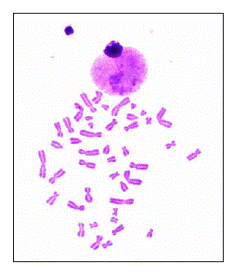 |
| 12- Comparative study between angiotensine converting enzyme inhibitor (Lisinopril) and angiotensine receptor blocker (Valsartan) on blood pressure and the incidence and severity of the side effects in hypertensive patients |
Kassim J. Al-Shamma*; Ibrahim Adham** and Musleh Ibrahim***
*Faculty of pharmacy and medical sciences, Al-Ahliyya Amman University, Amman, Jordan.
**College of pharmacy, university of Baghdad, Baghdad, Iraq.
***College of pharmacy, Hawler medical university, Iraq.
|
Abstract: Clinical observations and literature survey indicated the valsartan (angiotensine receptor blocker) has advantages over lisinopril (angiotensine receptor inhibitor) in controlling blood pressure and the incidence and severity of side effects in hypertensive patients. In this work, we explore this in 23 patients with essential hypertension treated with valsartan for 9 weeks, 24 patients with essential hypertension treated with lisinopril for the same period and 20 healthy normotensive individuals as control.
Blood pressures were recorded at 0,1,3,5,7 and 9 weeks after treatment ; blood samples were withdrawn from the patients and lipid profile parameters ( TC, TG, and HDL-C )were recorded at the same intervals ; sodium ion , potassium ion , urea and creatinine were also recorded at the same intervals. Follow up observations for the incidence and severity of dry cough were also recorded.
The results indicated that valsartan had significantly more hypotensive effect than lisinopril; moreover, valsartan had more lowering effect on total cholesterol than lisinopril. Patients on lisinopril treatment complained the presence of persistent dry cough, while those on valsartan treatment suffered only and rarely from mild dry cough.
|
| 13- Intestinal parasitoses in the students non permanent resident in Tunisia |
Introduction: The digestive parasitoses are affections which can be tolerated perfectly or on the contrary can determine serious disorders.
Objective: to evaluate the prevalence of the parasitoses in the non permanent resident students in Tunisia and to determine the principal parasitic species.
Material and methods: It is a retrospective study curried at the regional laboratory of hygiene of the CHU Hedi Chaker of Sfax during 12 years (1996 – 2007). This work has interested 4877 students non permanent resident. All specimens were examined by a direct examination and two methods of concentration.
Results: Our results show that 24.7
|
| 14- First identification of C.dubliniensis in Sfax- Tunisia |
Khlif Mohamed, Ben. Abdallah Fathia., Kanoun Amel., Sellami hayet, Sellami Amira, Cheikhrouhou
Fatma., Makni Fattouma, Ayadi Ali
Habib Bourguiba Hospital- Sfax Tunisia.
|
Introduction: Candida dubliniensis, a newly described fungal pathogen associated mainly to immunocompromised host’s infection is phenotypically closely related to C. albicans.
Objective: To determine the prevalence of C. dubliniensis from ICU HIV negative patients.
Patients and methods: prospective study from January 2004 to June 2004. 367 isolates identified by conventional methods as C. albicans obtained from 142 HIV negative patients hospitalized in the ICU at Habib Bourguiba Sfax Hospital. Our study was based on the failure to grow at 45°C, chlamydospore production and Colony morphology on Staib agar, colony color on Candiselect4 (Bio Rad) and the latex agglutination test. The confirmation of the discrimination between both species was performed by PCR targeting the hyphal wall protein 1 gene in the both species.respectively.
Results: 20 isolates were considered suspicious due to no growth at 45°C. The culture on Candiselect 4: C. albicans developed a pink colony and the C. dubliniensis developed a dark green colony on this media. Fourteen isolates were identified as C. dubliniensis. All the 20 isolates were grown on the Staib agar media, 14 of them presented rough colonies with hyphal fringes and have produced abundant chlamydospores. Of these 20 isolates, 14 gave a positive reaction with the agglutination test. The PCR has confirmed the identity of the 14 isolates as C. dubliniensis. The isolates of C. dubliniensis identified in this study were the same by the various tests applied. The prevalence of C. dubliniensis found in our clinical specimens was of 3.8
|
| 15- Ameliorating Serotypability of Pseudomonas Aeruginosa Strains in Immunocompromised Patients |
S. Khsiba, W. Achour, A. Ben Hassen*
*Laboratory of the Bone Marrow Transplant Center of Tunisia.
|
Pseudomonas aeruginosa is a common pathogen causing opportunistic infections particularly among immunocompromised patients and frequently implicated in nosocomial outbreaks. Serotyping is a simple and quick method frequently used for initial epidemiological studies. The aim of this study is to investigate the utility of 5 days subculture in serotyping of P. aeruginosa strains.
A total of 104 non replicate P. aeruginosa strains were isolated from clinical
specimen in 101 immuno-compromised patients in the National Bone Marrow Transplant Center of Tunisia from 2003 to 2007. Strains were identified by conventional methods and Api 20NE (BioMérieux). Serotyping was performed by agglutination on slides with commercially obtained antisera (Sanofi, Pasteur) at initial isolation and after 5 days of subculture on trypticase soja agar.
P. aeruginosa strains were isolated essentially from stools (52%) (colonisation), pus (16.5%), respiratory specimen (14,5%) and blood culture (9%). Serotypeability rate increased from 78% at initial isolation to 88.5% after 5 days of subculture. Serotyping resolved isolates especially into 5 serotypes: O:11 (32%), O:6 (21%), O:1 (16%), O:12 (10%) and O:10 (9%).
Twelve strains (11, 5%) were no serotypable.
In conclusion, a better serotypeability rate of P. aeruginosa strains was obtained after 5 days of subculture, ameliorating screening of outbreaks in our center.
|
| 16- Genetic Test and Counseling |
Lama Jabban
Genetic Disease Unit, Damascus University Children Hospital.
|
Genetic counseling is the process by which patients or relatives, at risk of an inherited disorder, are advised of the consequences and nature of the disorder, the probability of developing or transmitting it, and the options open to them in management and family planning in order to prevent, avoid or ameliorate it. This complex process can be seen from diagnostic and supportive aspects.
Specific tests may be requested following genetic counseling to confirm the diagnosis, plan for prenatal diagnosis, detect the carriers, or define affected presymptomatic individuals.
The genetic counselor must be aware of the psychological, moral, and social implications of the disease, and must take into due consideration the cultural background of the patient and family, without compromising the truthfulness of the scientific facts, or being obliged to hide any part of them.
Therefore, genetic consultation should help individuals and family translate scientific knowledge into practical information to understand genetic diseases and how inheritance works, and must support them to make personal decision about pregnancy, child care and genetic testing, but must not direct them to any particular alternative, or put them under any psychological pressure to reach a certain decision.
This lecture explains the scientific and moral dimensions of genetic testing and counseling and highlights important guidelines by presenting some complicated cases and discussing the impacts of the genetic consultation on the future of the individual and family.
|
| 17- Epidemiologic profile of bacteremia due to extended spectrum beta-lactamase producing enterobacteria
at Charles Nicolle hospital of Tunis
|
| Miled D., Mabrouka S. Mohamed Hamzaoui and Ben Redjeb S.
Laboratoire de microbiologie, boulevard 9 avril, Tunis, Tunisia
|
Extended spectrum beta-lactamases (ESBL) - enzymes which hydrolyze beta-lactam antibiotics, mainly third generation cephalosporins- are essentially described in Enterobacteriaceae. Infections due to pathogens producing such enzymes notably systemic ones can be life threatening because of their frequently associated resistances. The purpose of this 8 -year study (January 2000-december 2007) is to determine the epidemiologic profile of bacteremia due to ESBL producing enterobacteria (ESBL-PE).
Methods: Bacteriologic diagnosis of bacteremia was done by BacT/Alert automated system (bio-Mérieux®), bacterial identification was done by conventional methods, antibiotic susceptibilities were performed by disc diffusion method and ESBL detection by double synergy test.
Results: In this study, 323 ESBL-PE were isolated which represents 24.5
|
| |
| Mitra A. Tabari, Javad Shokry and Rashed F.
Shahid Beheshti Hospital, Laboratory
|
| Patient With: Liver Abscess in right Lobe, 18 years old, Thalasemia Major, Fever along 1 month.
Direct Smear: with many WBC.
Culture specimens on these media (EMB)(BLOOD AGAR)(CHOCOLATE AGAR)(SELENITE F) (TIOGLYCOLATE) (BISMOTE SULFATE (WILSON BELER) (XLD) (DXOXYCOLATE)
24 hour incubation in 37 centigrade.
Subculture from Selenite F on these media were mucouid colony.
On Bismote sulfate colony was black.
Differential panel from Bismote sulfate media were (Glu+) (Lac-) (Indol-) (Cimon Citrate+) (Metyle Red+) (Voges Proskaer-) (Motility+) [Lda + (Lyzin Deaminase)] [Ldc + (Lyzin Decarbocsilase)] (Bile Sculin +) [Odc + (Ornitin Decarbocsilase)]
Widal test titer for OD, OA were 1/40 positive.
Serological antiserum with colony was negative.
Attend to grow this bacteria on Selenite F, grow on Bismute Sulfate media in subculture and compraition biochemistry characterize, response to salmonella drug, admitted by microbiology ward of reference laboratory isolated salmonella Spp.
|
| 19- Place of Bone Markers in Evaluation of Osteoporosis Treatment |
| Mouna Dimassi |
| Description: The efficacy of osteoporosis treatment is usually evaluated on modifications of bone mineral density (BMD) after at least 2 years of therapy and on happening of atraumatic fracture. Many studies suggest the interest of the bone markers on earlier evaluation. The aims of our study were to determine the evolution of bone markers in osteoporosis with treatment.
Materials and methods :It was a prospective study including 148 postmenopausal women whose mean of age was 56 years (43-85 years).The first subgroup (GA) was constituted of 86 women with atraumatic fracture and receiving bisphosphonates (Résidronates 5 and 35 mg).The second group (GB) was composed of 62 women without osteoporotic fracture and receiving viatminocalcic supplementation. The measurements of bone formation markers (bone ALP) and osteoresorption markers (?-crosslaps or CTX) were performed before treatment, at 3 months and 6 months.
Results: Only 58.1
|
| 20- Diabetes mellitus and hemoglobinopathies: Which method to control glycated haemoglobin? |
| N.Ghrairi, K.Bouzid, A.Bahlous, A.Ben Khaled, M.Dimassi, E.Haouala, R.Baccar, J.Abdelmoula.
Biochemistry Department- Charles Nicolle Hospital; Tunis-Tunisia.
|
| Introduction: Glycated haemoglobin (HbA1C) is a marker used to monitor patients with diabetes mellitus.
It’s used to assess long-term glycemic control.
Hemoglobinopathies who are frequent diseases in our country affect the accuracy of HbA1C and leads to a wrong interpretation.
The aim of our study is to precise the optimal method in the assay of HbA1C when hemoglobinopathies are present.
Materials and methods: We have studied 200 samples of diabetic patients. The collection of whole blood was in EDTA tube and the HbA1C assay done simultaneously with two different methods: immunoturbidimetric assay on INTEGRA 400 (ROCHE) and ionic exchange high pressure liquid chromatographic assay (HPLC) on D-10 (BIO-RAD).
Results: Among the 200 plasmas four cases (2
|
| 21- Evaluation of the treatment response of genotype 1 chronic hepatitis C virus infections |
| Nahed Hogga, Selma Mhalla, Nejia Riahi, Amel Sadraoui, Olfa Bahri and Hinda Triki
Laboratory of Clinical Virology, Institut Pasteur of Tunis, Tunisia
|
| The hepatitis C is a major problem of public health because of its association with high frequency of chronicity and hepatic complications. The hepatitis C virus (HCV) is the agent responsible of this worldwide infection and includes at least 6 genotypes. Genotypes 1 and 4 are less responsive than the other genotypes to interferon therapy. In Tunisia, molecular studies reported the predominance of genotype 1. The aim of this work is to evaluate the virological response of this genotype to the bi-therapy. Seventy four patients infected with HCV of genotype 1, treated by biotherapy and followed during the period of 2002-2007, were investigated. For all the patients, pre-treatment viral quantification and genotyping were conducted by PCR-hybridization techniques, HCV Monitor-Roche and INNO LiPA-Innogenetics, respectively. In order to check the early virological response, HCV quantification was carried out after 12 weeks of treatment. A PCR of detection (HCV Amplicor – Roche) was conducted at the end of the treatment (1 year) for 38 patients and 6 months after, for 18 patients, in order to investigate the long-term virological response. A virological response at the end of the treatment was found in 71 |
| 22- Health Risks from Microbial Growth and Biofilms in Drinking Water Distribution Systems in Palestine
|
Description: Water distribution systems play a major role in determining the final quality of potable drinking water. Pathogenic and toxigenic microbiological agents in drinking water can cause diseases and death to consumers. The health risks associated with these pathogens range from viral and bacterial gastroenteric diseases to infections such as hepatitis A and giardiasis. Drinking water samples (n=24) with a volume of 1000 ml were collected in sterile bottles from the distribution systems at different regions in Ramallah District in Palestine. Moreover, swabs from the inside of the water distribution system of the same regions were taken. Sample were filtered through 0.45uM membrane, and various tests were conducted on each sample including total coliform, fecal coliform, heterotrophic plate count, Pseudomonas auroginosa count, fecal streptococci, sulfite reducing anaerobes, residual chlorine, turbidity, ammonium and nitrate. For protozoa, microscopic examination was done for the swabs transported in saline from the biofilms within the same day of sampling. Four samples out of 12 (33
Signal Transduction in Chronic Lymphocytic Leukemia B –Lymphocytes
The B-cell receptor (BCR) is a key signaling molecule that engages antigens and triggers pathways that induce survival, proliferation, differentiation, anergy, or apoptosis. The “choice” of which functional fate the B cell makes is decided upon by a series of receptor and cellular interactions. The potential for ligand-initiated signal transduction through the BCR is assessed in terms of ligand binding affinity for membrane Ig. We have used a series of anti-IgM H chain mAb that differ in their affinities (HB57 - Ka = 5x10⁸ "high", Mu53 - Ka = 2x10⁷ "intermediate", and P24 - Ka = ~2x10⁶ M-1 "low") in both soluble and immobilized forms as models for antigenic stimulation of the BCR, and assessed the response of normal B cells isolated by negative selection from peripheral blood. Proliferative responses were observed upon cross-linking the BCR with antibodies of all affinities at 10 ug/ml of immobilized antibodies for 72 hours, with a modest stimulation at 1ug/ml and no stimulation at either 0.01 or 0.1 ug/ml. Interestingly, the low affinity antibody P24 showed the highest proliferative response (3.3 fold), while stimulation with the moderate affinity antibody was intermediate (2.2 fold) and the response of the high affinity antibody, was negligible. Soluble antibodies did not stimulate proliferation at any concentration tested. When B cells were co-incubated with soluble anti-IgM antibodies plus IL 4,10ug/ml of either high or intermediate affinity induced proliferation with stimulation indices of 2.5 and 1.9, respectively, while the low affinity antibody still had no effect. Co-incubation with IL 4 and immobilized anti-IgM antibodies induced the same pattern of responses as seen with antibody alone, slightly increasing proliferation induced with 1 ug/ml low and intermediate and markedly increasing stimulation by all three antibodies at 10ug/ml. Our results suggest that even in the absence of T cell help, normal B cells can be activated via the BCR upon cross-linking and the efficiency at which this is accomplished is largely dependent on affinity for the BCR.
Key words: B cell, Signal transduction, Antibody response |
| 23- Renin-Angiotensin-Aldosterone System Haplotypes are Associated with Ischemic Stroke in Adult TunisianStroke Patients |
Saidi S.1, Bouassida S.2 and Mahjoub T.1
1Faculty of Pharmacy, University of Monastir, Monastir
2Arabian Gulf University, Manama, Bahrain.
|
| Objective. Insofar as the renin-angiotensin-aldosterone system (RAAS) contributes to stroke pathogenesis, brain infarction (BI), this study investigates the association between 4 different RAAS polymorphisms (renin C-4063T, and angiotensinogen T174M, M235T, and A-6G) and stroke. Subjects and Methods. Study subjects comprised 329 Tunisian Arab stroke patients (180 men and 149 women) and 444 age- and gender-matched controls (245 men and 199 women). Genotyping was done by PCR-RFLP analysis, and the contributions of RAAS polymorphisms to stroke were analyzed by haplotype analysis and multivariate regression analysis. Results. The frequencies of (mutant) AGT 235T and AGT -6G alleles, together with AGT 235T/T, AGT -6A/G and AGT -6G/G genotypes were higher in patients than in control group; the allele and genotype distribution of AGT T174M and renin C-4063T SNPs were comparable between patients and controls. Strong LD linkage disequilibrium was noted for AGT 174T with AGT 235M and with AGT -6A among patients only, while AGT 235M was in LD with AGT -6A in controls, and AGT 235T was in LD with AGT -6G in both patients and controls. The 174T/235T/-6A haplotype (P = 0.001) was positively, and the 174T/235M/-6G (P < 0.001) haplotype was negatively associated with stroke. Taking 174T/235M/-6G haplotype as reference, univariate analysis identified the 174T/235M/-6A (p = 0.001), 174T/235T/-6G (p = 0.001), 174T/235T/-6A (p < 0.001), and 174M/235T/-6A (p = 0.003) haplotype to be positively associated with stroke. Conclusions. These results suggest that RAAS polymorphism influence the risk of stroke in Tunisian Arab patients.
|
| 24- Polymorphisms of the Human Platelet Alloantigens
HPA-1, HPA-2, HPA-3, and HPA-4 in Ischemic Stroke
|
Saidi S.1 Mahjoub R.2 and Mahjoub T.1
1Faculty of Pharmacy, University of Monastir, Monastir
2Arabian Gulf University, Manama, Bahrain.
|
| Platelets regulate several aspects of primary hemostasis by maintaining vascular integrity, wound healing and repair at sites of injury, and by responding to endothelial damage. Altered platelet activation and platelet-dependent thromboembolism was associated with the pathogenesis of cerebrovascular diseases, including stroke. Insofar as the renin-angiotensin-aldosterone system (RAAS) contributes to stroke pathogenesis, brain infarction (BI), this study investigates the association between 4 different RAAS polymorphisms (renin C-4063T, and angiotensinogen T174M, M235T, and A-6G) and stroke. Polymorphism in human platelet antigen (HPA)-1 and HPA-3 (GPIIb/IIIa), HPA-2 (GPIb/IX), HPA-4 (GPIIIa) and HPA-5 (GPIa/IIa) was investigated in 216 stroke patients and 318 matched control subjects. HPA genotyping was done by PCR-SSP method. Lower HPA-1a (p < 0.001) and higher HPA-1b (p < 0.001) allele frequency were seen in patients than control subjects, and homozygosity for HPA-1b (p < 0.001) alleles was more prevalent in stroke cases than controls. The allele and genotype distributions of the other HPA polymorphic variants were similar between cases and controls. Select HPA haplotypes comprising the 2121 (Pc = 0.008) and 2221 (Pc = 0.018) haplotypes, which were positively associated, and the 1111 (Pc < 0.001) which was negatively associated with stroke, thereby conferring a disease susceptibility and protective nature to these haplotypes. Multivariate analysis confirmed the negative association of the 1111 (p < 0.001) and the positive association of the 2121 (p = 0.017) haplotypes with stroke, after adjustment for the a number of covariates. This is the first evidence demonstrating differential association of the common 4 HPA gene variants and specific HPA haplotypes with stroke. |
| 25- Screening for subtelomeric rearrangements in patients with developmental delay and/or idiopathic mental retardation using Fluorescence in situ hybridization (FISH) |
Sameh AL-ASSIL
Specialised Genetics Centre
|
| Chromosomal rearrangements involving the ends of chromosomes (telomeres) have emerged as an important cause of human genetic diseases in general and mental retardation in particular since 1992. Conventional cytogenetic analysis is the standard investigation for suspected chromosomal rearrangements, yet it cannot routinely detect rearrangements smaller than 3-4 megabases (Mb), and much larger abnormalities escape notice if they occur in regions where the banding pattern is not distinctive. Fluorescence in situ hybridisation (FISH) studies using multi-subtelomeric probes are used successfully to screen for subtelomeric rearrangements between patients with developmental delay and/or idiopathic mental retardation and their apparently normal parents when requested via the clinical geneticists. FISH-based methods still provide the gold standard for the detection of copy number changes and for balanced rearrangements. Since FISH-screening for multi-subtelomeric chromosomes is labour intensive and time consuming the pre-selection criteria have to be enforced. |
| |
Sara Gharib1 and Mitra Amooghli Tabari2
1Ophthalmologist, Babol, Shahid Beheshti Hostipal, OENT ward
2Ms of Medical Mycology, Shahid Beheshti Hostipal, Microbiology ward
|
Patient: men, 68 years old, Farmer, eye exudates, admitted eye clinic,
treatment with antibiotic (Ciprofloxacin).Pain and eye exudates were current. Ophthalmologist saw by Slit Lamp radial symptom in cornea conduct to fungal keratit. Sampling from cornea by means bistory were done Ophthalmologist.In direct smear from cornea not were seen any fungal agent.
Culture on Blood Agar, Chacolate Agar, Mac congy Agar, Tioglycolate and
Saboro Dexstrose Agar.Total media except Saboro Dexstrose Agar were incubation in 37 C for 24 hours .In this time no grow bacterial agent.In Subculture from Tioglycolate on Blood Agar, Chocolate agar, Macongy agar and incubation in 37 C for 24 hours no grow bacterial agent.After 10 days on Saboro DexstroseAgar in room temperature (25) and moisty grown colony cotton that first white color aftertime grows pink and red color.In direct smear from colony macroconidia banana form.Treatment with antifungal (Natamycin) is successful. Radial symptom disappear and reduce eye exudates and better patient vision. Notice to prevalence Fuzarium in field soil and occupation patient (farmer) this fungi is agent keratit published
|
| 27- Importance of confocal microscopy on dermatopathology |
| Toumi I; Jaafar S; Sriha B. and Korbi S. |
In this study we are interested to the morphologic investigation using confocal
microscopy (MCF). This technique is based on the fluorescence microscopy; light source is replaced by a laser beam. This instrument provides a very large spectrum of employment.
It is specially used for measuring dynamics process, and among different biological areas where it is used, we have proven its usefulness on dermatopathology especially in Bullous Dermatitis Autoimmune (DBAI), which generally requires an Immunofluorescence examination. In our study, we began to work on two types of DBAI widespread in Tunisia which are pemphigus and pemhigoide Bullosa, the antigenic targets in those disease are desmogline 1 and 3, present in the hemidesmosomes localized at the dermal-epidermal junction.
The MCF enable better morphological analysis of the immuno-fluorescence and gets good 2D detailed image with a collection of information from different optical sections needed to build 3D specimen. This makes it the most sophisticated instrument for certain diagnosis.
|
| 28- Optimisation of molecular techniques for the study of Dientamoeba fragilis |
| Trabelsi H; Cheikhrouhou F; Ben Abdallah F; Kanoun A; Sellami H; Sellami A; Makni F. and Ayadi A
Laboratoire de Parasitologie- mycologie Hal Habib Bourguiba - Sfax – Tunisia
|
| Dientamoeba fragilis is one of the parasites least studied protozoa. The majority of the authors estimate that D. fragilis is an important enteric pathogenic agent with an impact on the symptomatic infection varying from 4 to 91. |
| 29- Absence d'association entre les polymorphismes du gène de l’eNOS, le taux plasmatique de l'homocystéine et le risque des fausses couches répétées inexpliquées chez les femmes tunisiennes |
| Walid Zammiti, Nabil Mtiraoui et Touhami Mahjoub |
| Les polymorphismes du gène de l’eNOS ont été décrits associés avec une réduction de la production du monoxyde d’azote vasculaire qui est un agent vasodilatateur et antiagrégant plaquettaires. Le NO intervient aussi dans le catabolisme de l’homocystéine.
L’objectif de ce présent travail est d’évaluer l’effet des certains polymorphismes fonctionnels du gène de l’eNOS (894 G/T, -786 C/T et 4a/4b) et le risque des pertes récurrents de grossesses inexpliquées. Cette étude a été menée sur 350 patientes ayant au moins trois fausses couches inexpliquées et 200 témoins n’ayant aucuns antécédents d’avortements. La recherche des polymorphismes du gène de l’eNOS a été réalisée par PCR-RFLP, alors que le taux plasmatique de l’homocystéine est effectué par ELISA.
Les résultats obtenus montrent les fréquences allèliques, génotypiques et haplotypiques de tous les variants analysés sont comparables entre patientes et témoins. De même, le taux plasmatiques de l’homocystéine est similaire entre les deux groupes (p=0.06). Aucune variation du taux de l’homocystéine n’est démontrée en association avec les différents génotypes des polymorphismes du gène de l’eNOS. Il ressort de ce travail l’absence d’une association entre les polymorphismes -894G/T, -786C/T et 4a4b du gène de l’eNOS et le risque de fausses couches répétées ainsi que pour la variation du taux plasmatique de l’homocystéine
|
| 30- Toxoplasmic seroprevalence in the premarital assessment at the regional laboratory of hygiene CHU Hedi chaker Sfax. Tunisia |
Introduction: Rubella is a viral contagious infection generally occurring among the children between 5 and 9 years. It is transmitted by respiratory way (benign acquired rubella) or by transplacentary way (serious congenital rubella). The disease is generally unapparent. In the other cases, it is characterized by a moderated fever, cutaneous eruption, muscular and articular pains and cervical adenopathies.
Objective: To determine the seroprevalence of rubella for the women at the premarital assessment.
Material and methods: This retrospective study was done on 46597 women who come in consultation at the regional basic care health service for the premarital assessment. This study was carried during 15 years (1992 – 2006).
The research of the IgG antibodies was effected by the method ELISA.
Results and discussion: 82
|
| |
| المجلد 4 ,
العدد 9
، ربيع الأول 1429 – نيسان (إبريل) 2008 |
|
|
|
